Printing human organs with a bioprinter
More recently, the British magazine The Economist published an exciting article about a bioprinter that will be used to print human organs!
Surgeons who transplant human organs hope that one day they will be able to get all the organs needed for a transplant at the first request. Now the patient can spend several months, and possibly years, in anticipation of an organ from a suitable patient. During this time, his condition may worsen. He may even die. Thanks to artificial organs, it would be possible not only to alleviate the suffering of patients, but also to save human lives. Now, with the advent of the first commercial 3D bioprinter, this opportunity may become a reality.
The printer, which cost $ 200,000, was developed as a result of the cooperation of two companies: Organovo, which is located in San Diego and specializes in regenerative medicine, and the machine-building Invetech, located in Melbourne. One of the founders of Organovo, Gabor Forzhak, developed a prototype on which the new 3D printer is based. The first working printer models will soon be delivered to research teams who, like Dr. Forzhak, are exploring ways to create artificial tissues and organs. Currently, most of this work is done manually, using existing devices.
')
According to Keith Murphy, the director of Organovo, at first only simple tissues will be created, such as skin, muscles and small areas of blood vessels. However, immediately after the end of the test of test samples, the production of blood vessels for operations will begin, when it is necessary to “lay” new vessels for the movement of blood to bypass the damaged ones. After further research, it will be possible to produce more complex organs. Since machines are capable of printing networks of branched vessels, it would be possible, for example, to create networks of blood vessels necessary for supplying blood to such artificially produced organs like the liver, kidneys, and heart.

Organovo's 3D bio-printer uses the same principle of operation as “regular” 3D printers. 3D printers work in the same way as regular inkjet printers, but they print the model in three dimensions. Such printers spray polymer droplets that fuse together and then form a single structure. Thus, for each pass, the print head creates a small polymer line on the object. As a result, step by step, the subject takes on its final form. Cavities in a complex object are supported with the help of “scaffolding” of special water-soluble materials. These stages are washed out after the object is completely finished.
Researchers have found that a similar approach can be applied to biological materials! If you place tiny sections of cells next to each other, they begin to “fuse” together. A number of technologies are currently being studied that would allow the creation of human organs from individual cells, for example, the technology of “pumping up” muscle cells using small machines.
Despite the fact that the printing industry of human organs is in its infancy, scientists can already boast successful examples of creating human organs from scratch. So, in 2006, Anthony Atala, together with his colleagues from the Wake Forest Institute for Regenerative Medicine in North Carolina, USA, created bladders for seven patients. All of them are still functioning.
 The process of creating a bladder occurred as follows. Initially, the doctor took a tiny sample of the patient's bladder tissue (to prevent the newly created organ from being rejected by the immune system). Then, the resulting cells were applied to the biological bladder, which was a supporting base, having the shape of a bladder heated to the temperature of the human body. The applied cells began to grow and divide. After 6-8 weeks, the bladder was ready for implantation in a patient.
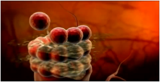
The advantage of using a bioprinter is that it doesn’t need a supporting framework (“scaffolding”). The Organovo machine uses stem cells derived from bone marrow. From stem cells, any other cells can be obtained using various growth factors. 10-30 thousand of such cells form into small droplets with a diameter of 100-500 microns. Such droplets retain their shape well and are great for printing.
So, the first printhead actually puts the droplets and cells in the right order. The second head is used for spraying a support base - a sugar-based hydrogel that does not interact and does not adhere to the cells. As soon as printing is completed, the resulting structure is left for one to two days in order for the drops to “fuse” with each other. To create tubular structures, such as blood vessels, a hydrogel is first applied (inside and outside the future structure). After that cells are added. As soon as an organ is formed, the hydrogel is removed from the outer part (like an orange peel) and pulled out of the inner part, like a piece of rope.
Other types of cells and supporting bases can be used in bioprinter. Thus, according to Mr. Murphy, liver cells can be applied on a preformed base, having the shape of a liver, or layers of connective tissue can be formed to create a tooth. At the same time, the new printer has such modest dimensions that it can be safely placed in a biological cabinet to provide a sterile environment during the printing process.

Some researchers believe that such machines as this one will ever be able to print tissues and organs right in the human body! And, in fact, Dr. Atala is now working on a printer that, after scanning a part of the body where skin grafting is needed, will be able to print the skin directly on the human body! Regarding larger bodies, Dr. Forzhak believes that they can take various forms, at least at the beginning. For example, in order to purify blood, an artificial kidney does not necessarily have to look like a real kidney or functionally repeat it completely. Those people who are waiting for organs will probably not be very worried about how new organs will look. The main thing is that they work, and people feel better.
PS Some of the processes described in the article, such as the formation of organs from cell droplets, are shown in the video:
Surgeons who transplant human organs hope that one day they will be able to get all the organs needed for a transplant at the first request. Now the patient can spend several months, and possibly years, in anticipation of an organ from a suitable patient. During this time, his condition may worsen. He may even die. Thanks to artificial organs, it would be possible not only to alleviate the suffering of patients, but also to save human lives. Now, with the advent of the first commercial 3D bioprinter, this opportunity may become a reality.
Creating a bioprinter
The printer, which cost $ 200,000, was developed as a result of the cooperation of two companies: Organovo, which is located in San Diego and specializes in regenerative medicine, and the machine-building Invetech, located in Melbourne. One of the founders of Organovo, Gabor Forzhak, developed a prototype on which the new 3D printer is based. The first working printer models will soon be delivered to research teams who, like Dr. Forzhak, are exploring ways to create artificial tissues and organs. Currently, most of this work is done manually, using existing devices.
')
According to Keith Murphy, the director of Organovo, at first only simple tissues will be created, such as skin, muscles and small areas of blood vessels. However, immediately after the end of the test of test samples, the production of blood vessels for operations will begin, when it is necessary to “lay” new vessels for the movement of blood to bypass the damaged ones. After further research, it will be possible to produce more complex organs. Since machines are capable of printing networks of branched vessels, it would be possible, for example, to create networks of blood vessels necessary for supplying blood to such artificially produced organs like the liver, kidneys, and heart.
The history of bio printing
Organovo's 3D bio-printer uses the same principle of operation as “regular” 3D printers. 3D printers work in the same way as regular inkjet printers, but they print the model in three dimensions. Such printers spray polymer droplets that fuse together and then form a single structure. Thus, for each pass, the print head creates a small polymer line on the object. As a result, step by step, the subject takes on its final form. Cavities in a complex object are supported with the help of “scaffolding” of special water-soluble materials. These stages are washed out after the object is completely finished.
Researchers have found that a similar approach can be applied to biological materials! If you place tiny sections of cells next to each other, they begin to “fuse” together. A number of technologies are currently being studied that would allow the creation of human organs from individual cells, for example, the technology of “pumping up” muscle cells using small machines.
Despite the fact that the printing industry of human organs is in its infancy, scientists can already boast successful examples of creating human organs from scratch. So, in 2006, Anthony Atala, together with his colleagues from the Wake Forest Institute for Regenerative Medicine in North Carolina, USA, created bladders for seven patients. All of them are still functioning.
The process of creating a bladder occurred as follows. Initially, the doctor took a tiny sample of the patient's bladder tissue (to prevent the newly created organ from being rejected by the immune system). Then, the resulting cells were applied to the biological bladder, which was a supporting base, having the shape of a bladder heated to the temperature of the human body. The applied cells began to grow and divide. After 6-8 weeks, the bladder was ready for implantation in a patient.The advantage of using a bioprinter is that it doesn’t need a supporting framework (“scaffolding”). The Organovo machine uses stem cells derived from bone marrow. From stem cells, any other cells can be obtained using various growth factors. 10-30 thousand of such cells form into small droplets with a diameter of 100-500 microns. Such droplets retain their shape well and are great for printing.
So, the first printhead actually puts the droplets and cells in the right order. The second head is used for spraying a support base - a sugar-based hydrogel that does not interact and does not adhere to the cells. As soon as printing is completed, the resulting structure is left for one to two days in order for the drops to “fuse” with each other. To create tubular structures, such as blood vessels, a hydrogel is first applied (inside and outside the future structure). After that cells are added. As soon as an organ is formed, the hydrogel is removed from the outer part (like an orange peel) and pulled out of the inner part, like a piece of rope.
Other types of cells and supporting bases can be used in bioprinter. Thus, according to Mr. Murphy, liver cells can be applied on a preformed base, having the shape of a liver, or layers of connective tissue can be formed to create a tooth. At the same time, the new printer has such modest dimensions that it can be safely placed in a biological cabinet to provide a sterile environment during the printing process.
Some researchers believe that such machines as this one will ever be able to print tissues and organs right in the human body! And, in fact, Dr. Atala is now working on a printer that, after scanning a part of the body where skin grafting is needed, will be able to print the skin directly on the human body! Regarding larger bodies, Dr. Forzhak believes that they can take various forms, at least at the beginning. For example, in order to purify blood, an artificial kidney does not necessarily have to look like a real kidney or functionally repeat it completely. Those people who are waiting for organs will probably not be very worried about how new organs will look. The main thing is that they work, and people feel better.
PS Some of the processes described in the article, such as the formation of organs from cell droplets, are shown in the video:
Source: https://habr.com/ru/post/89748/
All Articles