Simulation of view. Part one. Eye Excursion
1 Excursion in the eye - 2 Perception - 3 Geometry of view - 4 Eye tracking - 5 How to catch the eye - 6 Simulation of eye tracking
To understand how a person perceives an image, one will have to start from the organ of sight - from the eye. An important point for further understanding in addition to the anatomical structure of the eye is the limitation of the resolution of the eye, which I will describe here. If you know all this, then you can only briefly review the selected pieces of text and immediately go to the second part.
And the way the eye works, you probably remember everything from a textbook on biology, but here I will tell you some really amazing things that for some reason do not tell at school. But first, I will remind you of the eye device (illustrations are taken from David Hubel’s book Eye, Brain, Vision):
')
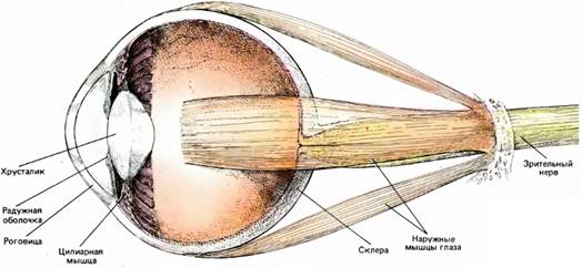
The eye is held in the eye socket by a group of 6 muscles, and they turn it up and down right and left, if necessary, “squint eyes”.
The luminous flux entering the retina passes through the cornea (it provides about 70% of light refraction), then through the pupil, which is analogous to the camera diaphragm and controlled by a group of radial and annular muscles that change its size, then enters the lens, which provides the final focusing visible objects. The lens is a gelatinous pad, which is compressed by radial muscles. During compression, the lens changes its shape, thus changing the degree of light refraction and focal distance.
Actually, all this is necessary to create a projection of the visible world on the retina. The retina is a part of the brain that separated from it in the early stages of development, but is tightly connected with it by the bundle of the optic nerve:
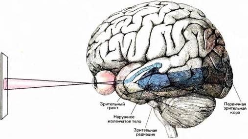
The retina is not just an analogue of a camera's sensor that converts light signals into electrical impulses. The retina performs the initial processing of the incoming image, before it enters the visual cortex of the brain.
The retina itself consists of three layers of nerve cells, and the actual photosensors that perceive light (rods and cones) make up the third, outer layer on its back surface:

Thus, in order to reach the sensors, the light first passes through two layers of nerve cells. The rear side photoreceptors are coated with melanin (black pigment), which plays the same role as blackening the inner body of the camera. If there was no blackening, the light, passing through a layer of rods and cones, would go farther to the brain, reflect from it and come back, in every way spoiling our picture of the world.
Photoreceptors are of two types:
Since the conversation is about analyzing images on a computer monitor, I will not talk more about scotopic vision and wands, focusing all my attention on photopic and cones.
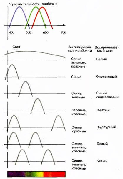
So, cones are divided into three types by the type of pigment in them, and each of these types is responsible for the perception of its spectral band. Conventionally, the types of cones are called "blue", "green" and "red", but in fact the spectrum of their perception goes beyond the specified colors. Therefore, many parts of the spectrum, for example, yellow, are perceived by two types of cones at once, and the cumulative sensation of excitement of the "red" and "green" cones creates in us a feeling of yellow color .
(more accurate graphs of the sensitivity distribution of cones can be found here )
There are approximately the same number of cones of all three types, but since in the area of the yellow spectrum the number of cones perceiving it is twice as large, the yellow seems much more intense than blue . The same applies to some other colors (see the diagram below).
All photoreceptors are combined by ganglion cells into blocks called receptive fields — each receptive field is assigned to each ganglion cell. One photoreceptor can consist in several receptive fields at once, therefore the fields of two adjacent ganglion cells overlap by 70-80%. In the simplest case, the receptive field is similar to the analogue of a pixel of a photosensitive matrix in a camera. But not everything is so simple!
First, receptive fields differ in size depending on the area of the retina : for example, in the area of the eye fossa, the area of the retina, the corresponding area of greatest visual acuity, the receptive field is 1-2 mm (which corresponds to 2-3 angular minutes), - already up to 5 mm!
Secondly, the “pixels” of receptive fields differ from the pixels of the camera matrix in that there is a separation among them — some react to “on”, others on “off”. Those. Some fields respond exclusively to changes in light from darker to lighter, others - vice versa .
Thirdly, the fields differ in size depending on their types (conventionally, they denote P fields and M fields). M fields are larger, have a high reaction rate, but because of their size, the picture transmitted by them will be in a much worse resolution than the picture transmitted by the P fields. P fields are smaller, so the picture, which they convey is more accurate, but their reaction rate is not great. Thus, M fields capture movement and answer the question “where is the object (motion perception and depth)”, and P fields deal with color, shape and details of the visual display or answer the question “what are the objects (color vision and sharpness)” . With mild contrast, M fields deal, and P with sharp contrast. The longer the view remains in place, the more the role of the P fields increases.
Fourthly, not everything is so simple with these “pixels”: if the pixels of the photomatrix fix only the color change, the receptive fields are also able to react to lines, stripes, various rectangular segments with clear edges! Those. in addition to the information that there is a change in color / illumination, these “pumped pixels” also transmit information that there is a straight line, information about its length (so long that it goes beyond the limits of vision or the end of the segment is visible) and even about the direction ( tilt angle) given straight line !
1 Excursion in the eye - 2 Perception - 3 Geometry of view - 4 Eye tracking - 5 How to catch the eye - 6 Simulation of eye tracking
To understand how a person perceives an image, one will have to start from the organ of sight - from the eye. An important point for further understanding in addition to the anatomical structure of the eye is the limitation of the resolution of the eye, which I will describe here. If you know all this, then you can only briefly review the selected pieces of text and immediately go to the second part.
Eye anatomy
And the way the eye works, you probably remember everything from a textbook on biology, but here I will tell you some really amazing things that for some reason do not tell at school. But first, I will remind you of the eye device (illustrations are taken from David Hubel’s book Eye, Brain, Vision):
')
The eye is held in the eye socket by a group of 6 muscles, and they turn it up and down right and left, if necessary, “squint eyes”.
The luminous flux entering the retina passes through the cornea (it provides about 70% of light refraction), then through the pupil, which is analogous to the camera diaphragm and controlled by a group of radial and annular muscles that change its size, then enters the lens, which provides the final focusing visible objects. The lens is a gelatinous pad, which is compressed by radial muscles. During compression, the lens changes its shape, thus changing the degree of light refraction and focal distance.
Actually, all this is necessary to create a projection of the visible world on the retina. The retina is a part of the brain that separated from it in the early stages of development, but is tightly connected with it by the bundle of the optic nerve:
The retina is not just an analogue of a camera's sensor that converts light signals into electrical impulses. The retina performs the initial processing of the incoming image, before it enters the visual cortex of the brain.
The retina itself consists of three layers of nerve cells, and the actual photosensors that perceive light (rods and cones) make up the third, outer layer on its back surface:
Thus, in order to reach the sensors, the light first passes through two layers of nerve cells. The rear side photoreceptors are coated with melanin (black pigment), which plays the same role as blackening the inner body of the camera. If there was no blackening, the light, passing through a layer of rods and cones, would go farther to the brain, reflect from it and come back, in every way spoiling our picture of the world.
Photoreceptors
Photoreceptors are of two types:
- The rods are receptors that are sensitive to very low light (mainly in the yellow-green part of the spectrum) and perceive very thin light variations, therefore, at high light levels, they become useless and are responsible for night, scotophous vision;
- Cones are receptors, for which work requires much more light, they perceive stronger gradations in illumination and are responsible for daylight, photopic vision.
Since the conversation is about analyzing images on a computer monitor, I will not talk more about scotopic vision and wands, focusing all my attention on photopic and cones.
So, cones are divided into three types by the type of pigment in them, and each of these types is responsible for the perception of its spectral band. Conventionally, the types of cones are called "blue", "green" and "red", but in fact the spectrum of their perception goes beyond the specified colors. Therefore, many parts of the spectrum, for example, yellow, are perceived by two types of cones at once, and the cumulative sensation of excitement of the "red" and "green" cones creates in us a feeling of yellow color .
(more accurate graphs of the sensitivity distribution of cones can be found here )
There are approximately the same number of cones of all three types, but since in the area of the yellow spectrum the number of cones perceiving it is twice as large, the yellow seems much more intense than blue . The same applies to some other colors (see the diagram below).
Retina Pixels
All photoreceptors are combined by ganglion cells into blocks called receptive fields — each receptive field is assigned to each ganglion cell. One photoreceptor can consist in several receptive fields at once, therefore the fields of two adjacent ganglion cells overlap by 70-80%. In the simplest case, the receptive field is similar to the analogue of a pixel of a photosensitive matrix in a camera. But not everything is so simple!
First, receptive fields differ in size depending on the area of the retina : for example, in the area of the eye fossa, the area of the retina, the corresponding area of greatest visual acuity, the receptive field is 1-2 mm (which corresponds to 2-3 angular minutes), - already up to 5 mm!
Secondly, the “pixels” of receptive fields differ from the pixels of the camera matrix in that there is a separation among them — some react to “on”, others on “off”. Those. Some fields respond exclusively to changes in light from darker to lighter, others - vice versa .
Thirdly, the fields differ in size depending on their types (conventionally, they denote P fields and M fields). M fields are larger, have a high reaction rate, but because of their size, the picture transmitted by them will be in a much worse resolution than the picture transmitted by the P fields. P fields are smaller, so the picture, which they convey is more accurate, but their reaction rate is not great. Thus, M fields capture movement and answer the question “where is the object (motion perception and depth)”, and P fields deal with color, shape and details of the visual display or answer the question “what are the objects (color vision and sharpness)” . With mild contrast, M fields deal, and P with sharp contrast. The longer the view remains in place, the more the role of the P fields increases.
Fourthly, not everything is so simple with these “pixels”: if the pixels of the photomatrix fix only the color change, the receptive fields are also able to react to lines, stripes, various rectangular segments with clear edges! Those. in addition to the information that there is a change in color / illumination, these “pumped pixels” also transmit information that there is a straight line, information about its length (so long that it goes beyond the limits of vision or the end of the segment is visible) and even about the direction ( tilt angle) given straight line !
1 Excursion in the eye - 2 Perception - 3 Geometry of view - 4 Eye tracking - 5 How to catch the eye - 6 Simulation of eye tracking
Source: https://habr.com/ru/post/57679/
All Articles