Harm for the good: the immune system of the lamprey in the fight against human brain cancer

Our brain is our everything. The disruption of the work of this vital organ leads to terrible and sometimes fatal consequences. The complexity of the brain and its neural organization is enormous, which greatly complicates the process of treating a particular disease. As a rule, when we treat something, we try to get rid of the defects that the disease causes. But, what if you use these defects to combat what creates them? This is exactly what the authors of the research we are considering today decided to do. How did the scientists apply the disruption of the hemato-encephalic barrier, why do they need access to the extracellular matrix of the brain and what role did the lamprey fish parasite play in this? This will tell us the report of the research group. Go.
A bit of theory
First of all, it is necessary to deal with the characters in this laboratory play.
')
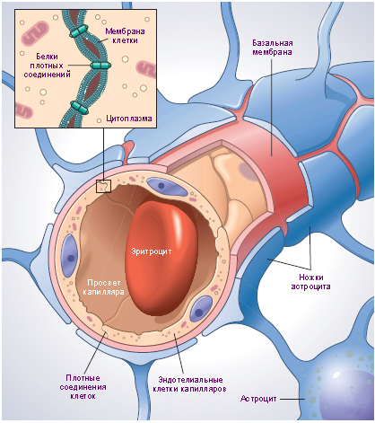
Hemato-encephalic barrier
One of the main roles is performed by the hemato-encephalic barrier (BBB) - a physiological barrier between the central nervous system (CNS) and the circulatory system. This barrier prevents the contact of nerve tissues with various components of the circulating blood, among which may be toxins, microorganisms, cellular / humoral factors of the immune system, which can respond to brain cells as if they were foreign. The BBB can be compared to a bouncer in a very expensive club, passing only nutrients into the central nervous system. But this bouncer is not particularly legible, he often does not miss the medications necessary for the treatment of the central nervous system. It turns out that a system aimed at the benefit of our health can be an obstacle to its treatment. So much for the irony in physiology.
However, the hemato-encephalic barrier does not always work like a Swiss watch. In cases of stroke, tumors, various head injuries, chronic diseases, the BBB begins to fail, that is, skip to the central nervous system what he would have previously weed out. In addition to the natural causes of failure, there is also an anthropogenic one: focused high-intensity ultrasound and osmotic agents that disrupt the BBB. Why break something that ensures the normal operation of the brain, you ask. Then, in order to deliver drugs that the full BBB will filter out. All the same, that the bouncer will not let the doctor in the club for a visitor who has fainted, because he does not have a club card.
The main and common for all cases, the result of the violation of the BBB is a pathological exposure of the extracellular matrix (ECM) of the brain, which is isolated in normal conditions.
Extracellular matrix is the basis of connective tissue, providing mechanical support for cells and transport of chemicals.
Therefore, scientists believe, aiming at certain parts of the brain with pathologically impaired BBB, it is possible to deliver drugs to those parts of the damaged CNS that were previously inaccessible because of the BBB.
Thus, you can create a ligand aimed at ECM, which will be effective in combating various diseases of the central nervous system, and not with one specific, as previously developed methods.
To test the theory in practice, the scientists decided to apply their method of drug delivery to incurable glioblastoma (brain cancer). This type of disease is quite rare, but it is extremely difficult to beat. Even after chemotherapy, radiation therapy and surgery, the survival rate is about 1-2 years.
Recent studies have shown that the use of immunotherapy with interleukin-13 * -related chimeric antigen receptors * is a fairly promising treatment for glioblastoma.
Interleukins * - peptide informational molecules produced by leukocytes, to a lesser extent, by phagocytes and other tissues. Interleukins are part of the immune system.
Interleukin-13 * (IL13) is the main mediator of physiological changes caused by allergic inflammation in many tissues.
Chimeric antigen receptor * is a recombinant fusion protein that binds an antibody fragment, is capable of selectively binding to specific antigens, and signaling domains that activate T-cells.The use of chimeric antigen receptors targeting the CSPG4 protein can also be a rather effective method for fighting glioblastoma.
In addition, MRI analysis showed a violation of the hemato-encephalic barrier inside glioblastoma. Therefore, treatment methods associated with the extracellular matrix should be effective.
And here comes the immense imagination and creativity of scientists. The point is that standard peptides and antibodies can be used as ECM-targeted reagents, but this is not so much fun. Therefore, scientists have decided to use variable lymphocyte receptors (VLR), that is, the antigen receptors of the lamprey.
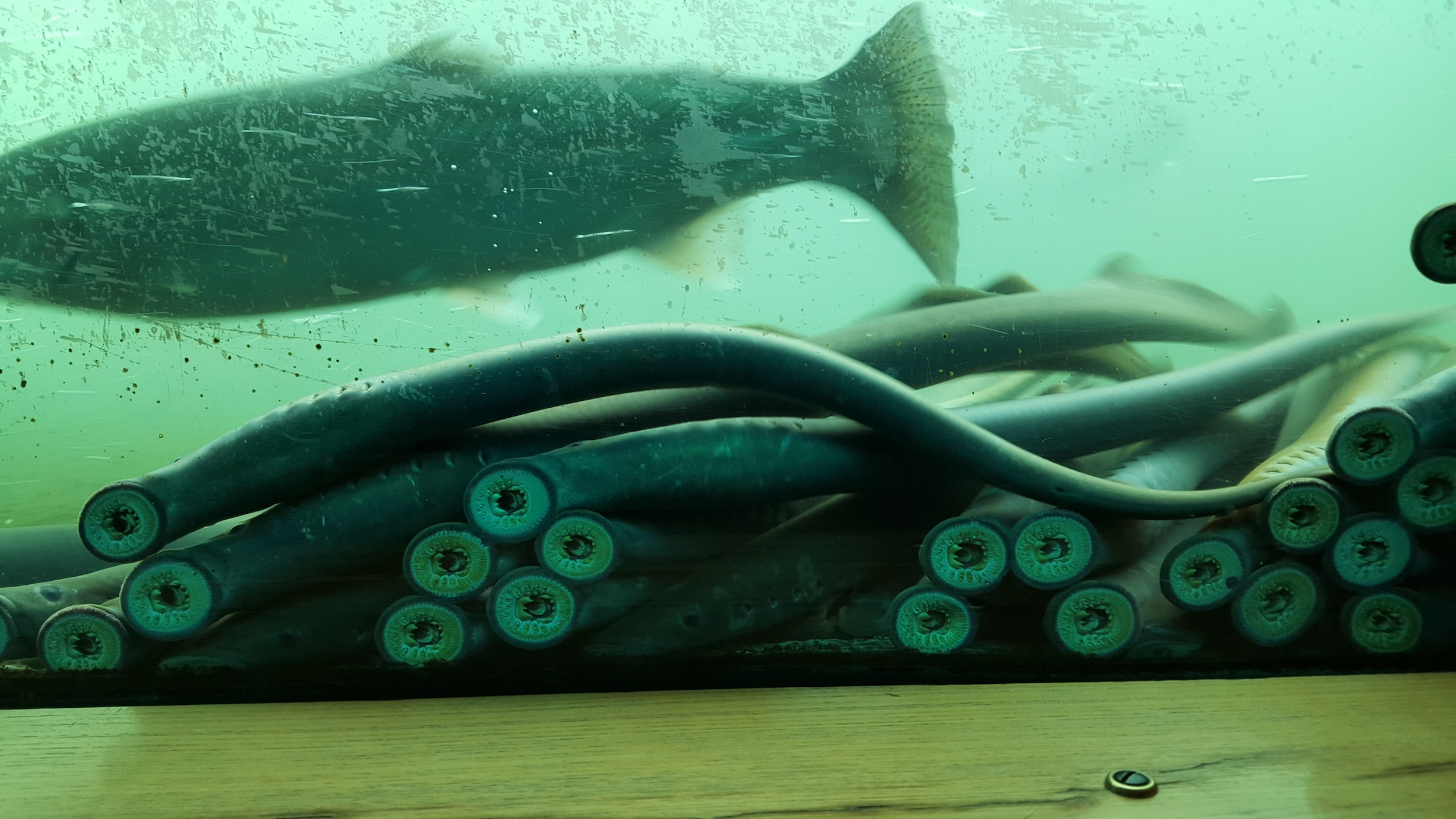
The class of lamprey has about 40 species, most of which are parasites that feed on the blood of fish to which they stick.
VLR are sickle-rich, leucine-rich proteins that recognize antigenic targets with specificity and affinity * , comparable to antibodies based on immunoglobulin.
Affinity * is a thermodynamic characteristic of the strength of the interaction of substances such as antigen and antibody.Why precisely lampreys and their VLR? The fact is that there is an evolutionary abyss of 500 million years between mammals and lamprey. Thus, lamprey VLRs are more likely to recognize conserved proteins and glycans in the ECM than mammalian antibodies.
To identify the VLRs that bind the ECM of the brain, scientists conducted a panning - a method of selecting certain bio-elements (proteins, petites, etc.) from the VLR libraries of biomolecules. The result was a pool of ECM-binding clones * .
A clone * is a group of identical cells that share a common ancestor (primary source), that is, they originate from the same cell.The resulting clone showed predominant accumulation in the destroyed areas of the hemato-encephalic barrier in animals with an osmotic violation of the BBB or glioblastoma. In addition, the clone perfectly targeted liposomes * loaded with doxorubicin (an antibiotic).
Liposomes * - spherical intracellular organelles used to deliver drugs to certain tissues.
Preparation for the study
As we learned earlier, ECM-binding VLRs have been identified through the panning of VLR libraries. The library itself was obtained from a combination of VLR lamprey immunized with mechanically isolated preparations of the plasma membrane of mouse microvessels, which contained the associated ECM of the brain.
The library was primarily enriched with ECM-binding using two panning cycles on a decellularized * ECM generated by cultured mouse brain endothelial cells (cell line bEnd.3).
Decellularization * is a method for cleaning allografts from the cellular component to obtain a non-immunogenic, efficient and safe construction based on the natural extracellular matrix.Further, it was necessary to identify precisely those ECM-binding clones, which predominantly bind bEnd.3 ECM, and not with the control group of mouse fibroblasts ECM (3T3 cell line).
Next, individual clones were placed in 96-well plates, after which they were expanded and induced to display the VLR. After removing the extra clones (to avoid subcloning), scientists were able to carry out a comparative assessment of VLR binding to bEnd.3 and 3T3 ECM using an ELISA screening ( 1a ).
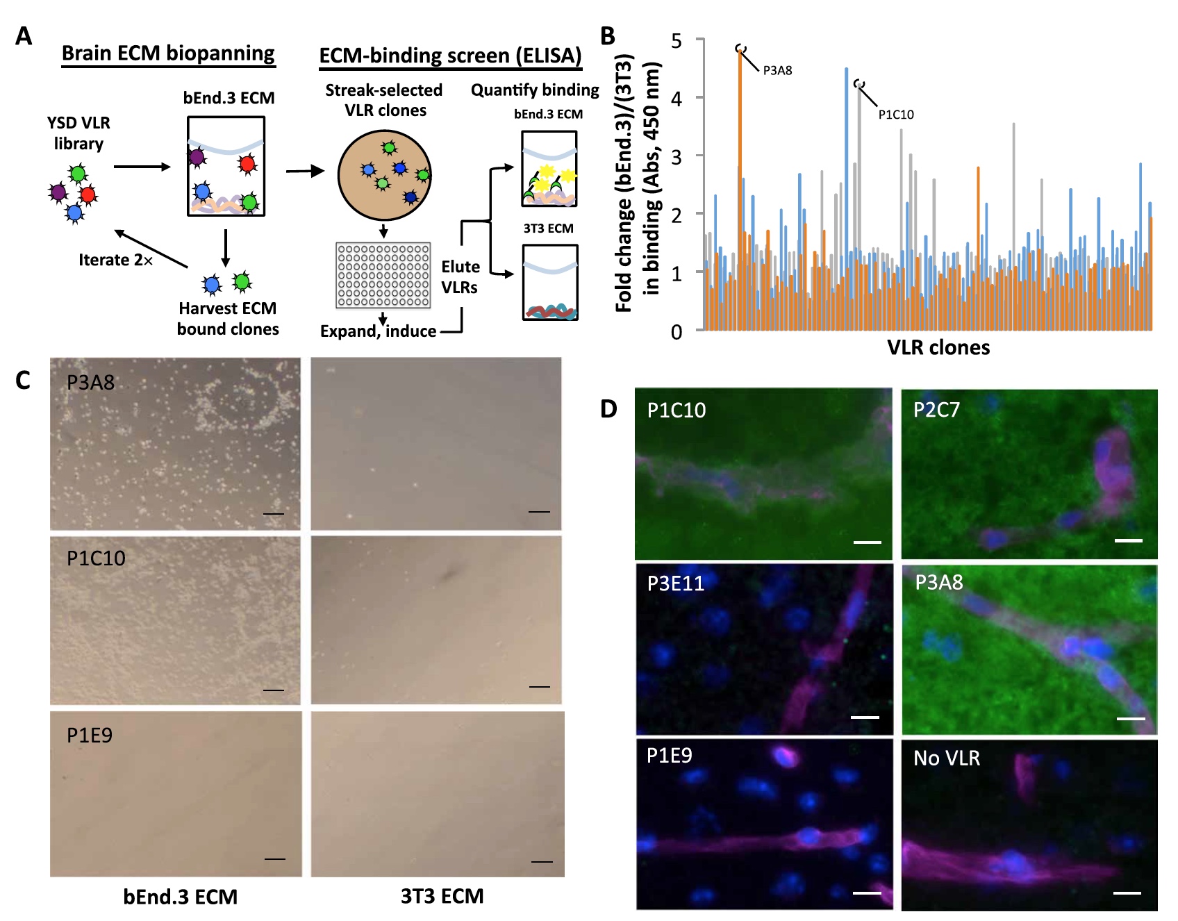
Image number 1
A total of 285 clones were analyzed. As a result, it is clear that communication signals with bEnd.3 ECM are about 5 times stronger than communication signals with 3T3 ECM ( 1b ).
Next, the ELISA results were checked by comparing the images of the bright field microscopy of clones associated with bEnd.3 and 3T3 ECM ( 1c ).
As seen in Figure 1c , clones P1C10 and P2C7 are associated exclusively with bEnd.3 ECM, and the non-binding clone P1E9 practically does not show any connection with any type of ECM.
Next, scientists conducted a comparative analysis of a more practical method - on the mouse brain. Eight of the 10 VLR clones that showed the best binding outcome in previous observations also demonstrated binding ( 1d ) in this analysis.
All binding clones showed a diffuse parenchymal EMC scheme without any additional enrichment (vascular or cellular).
Scientists identified 2 leaders on the results of all the above observations - P1C10 and P3A8. It is these clones that will be considered later.
Research results
P1C10 and P3A8 were functionalized with Cy5 fluorescent dye. Direct immunostaining of mouse tissues using VLR-Cy5 conjugates showed that P1C10-Cy5 has a significant selectivity towards the ECM of the brain compared to the kidney, heart and liver tissues ( 2a ).

Image number 2a
Immunostaining * is a process that allows you to identify and localize an antigen in a specific area of a cell, tissue, or organ.But P3A8-Cy5 binds to the ECM of the brain and liver with the same intensity, but just like P1C10-Cy5 shows no interest in the ECM of the kidneys and the heart.
Next, the scientists checked the cross-reactivity of P1C10 and the ECM of the human brain (cryo-slices were used). The binding of P1C10-Cy5 to the ECM of the human brain resembles a picture of the same process, but with the participation of the mouse brain ( 2b ). Also, P1C10-Cy5 successfully contacted the ECM in cryoprocesses of a sample of human glioblastoma ( 2c ).
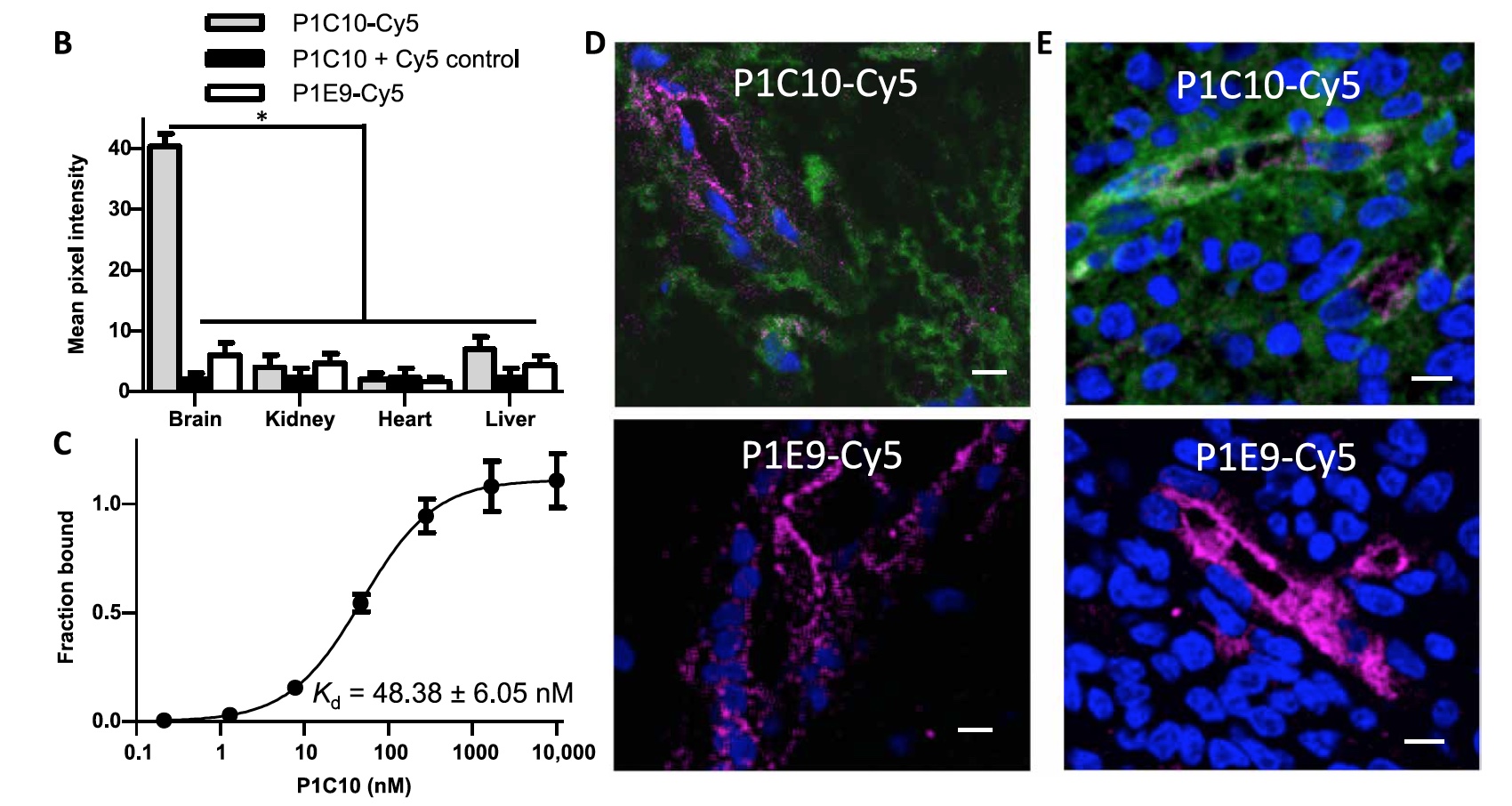
Image №2b-e
Given these observations, scientists measured affinity * P1C10.
Affinity * is the ability of a cell to capture and bind certain chemicals.As a result, the dissociation constant (Kd) for binding to bEnd.3 ECM was 48.38 ± 6.05 nM ( 2d ).
Following this, the scientists decided to check whether P1C10 will accumulate in the places of BBB destruction in the mouse brain.
Modified by the dye near-infrared radiation IR800 clones P1C10 or RBC36 were administered to healthy laboratory mice in a volume of 1 mg / kg. RBC36 is a VLR that recognizes the human H antigen trisaccharide, therefore it was used as an isotype control.
Next, mannitol (hexahydol alcohol) was injected intravenously into the mice for temporary disclosure of the BBB. After that, mice brain images were taken to identify the IR800 signals (image below).

Image number 3
A comparative analysis showed that the accumulation of fluorescence in the brain (concentration of the analyte, marked with IR800 dye) when using P1C10-IR800 is 3.3 times higher than with RBC36-IR800, and 7.6 times higher than when using saline. Consequently, P1C10 selectively accumulates in the brain after disruption of the blood-brain barrier.
Next, the scientists decided to check whether the VLR will target the exposed extracellular matrix. For this, two models were created using mouse GL261 and human U87 glioblastoma cells, which were introduced into the brain of experimental mice. As a result, tumors were formed with a chaotic vascular network and point violations of the hemato-encephalic barrier.
P1C10 or RBC36 in a volume of 1 mg / kg was administered intravenously to mice with inserted GB261 glioblastoma. After 30 minutes, brain samples were taken for analysis and visualization of the IR800 signal (dye for P1C10 or RBC36).
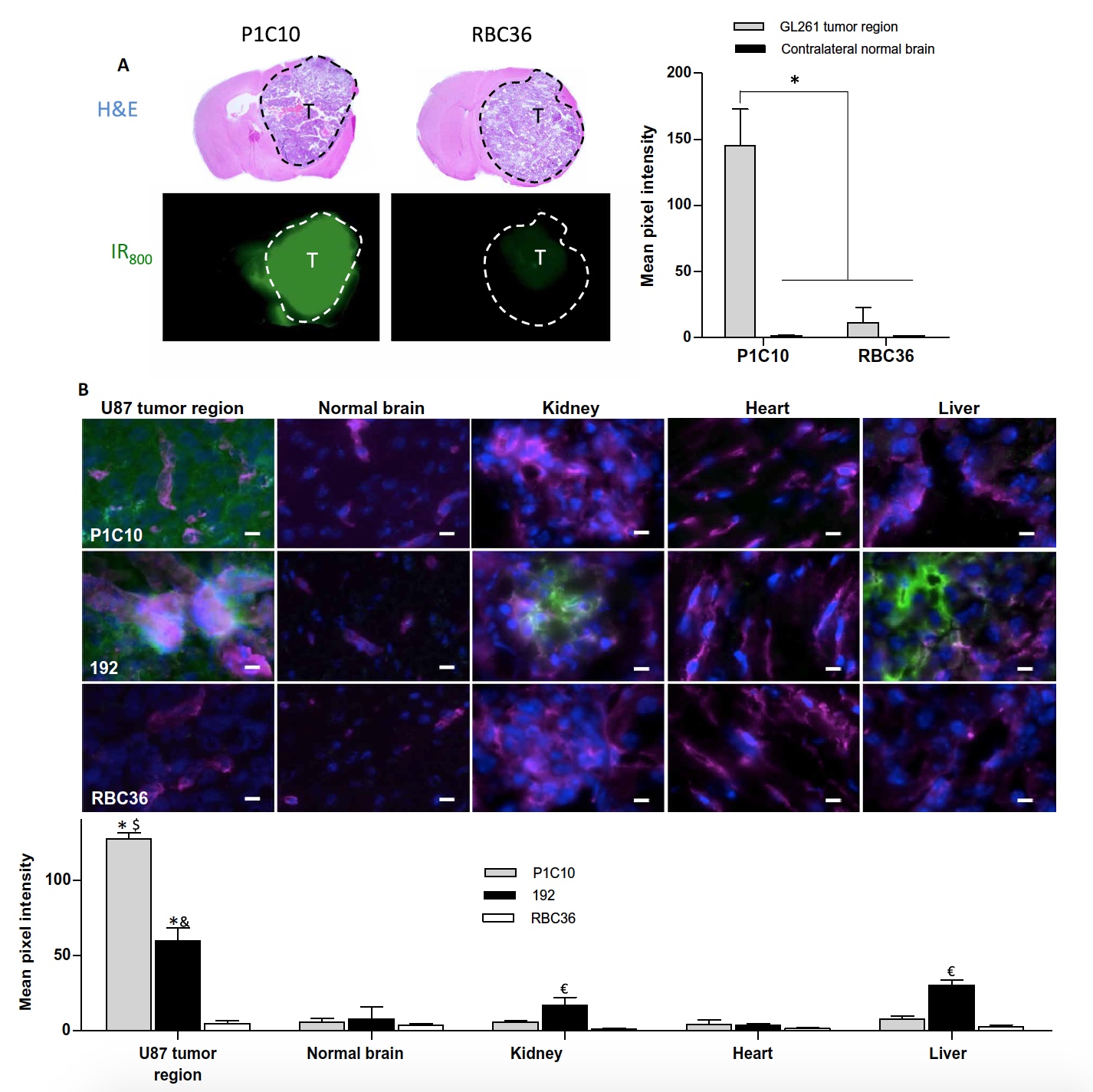
Image number 4
The average fluorescence intensity in the GL261 tumor region in mice that were injected with P1C10-IR800 was 112 times higher than in the contralateral (opposite) brain region ( 4a ). But when using RBC36-IR800, the fluorescence intensity of the tumor area was only 9 times higher than the intensity in the opposite area.
In addition, it was found that the accumulation of P1C10-IR800 in the tumor itself is 13 times higher than the accumulation of RBC36-IR800.
These observations confirmed the ability of P1C10 to selectively target ECM in mouse tumors. Now it was necessary to test this talent on human brain tumors.
VLR was administered to mice with U87 (a tumor of the human brain). Scientists tested not only P1C10, but also 192, which demonstrated selective binding with the basolateral side of the cerebral vascular network in addition to the vessels of the kidneys and liver, as well as to ECM.
P1C10, 192 or RBC36 in a volume of 3 mg / kg was administered intravenously and allowed to circulate freely for 30 minutes. After that, samples of organs of interest were taken to visualize the results.
Both VLR clones (P1C10 and 192) showed accumulation within the tumor, with P1C10 being distributed throughout the ECM of the tumor ( 4b ). But 192 for the most part concentrated outside the large tumor vessels.
None of the VLR clones accumulated in the healthy contralateral hemisphere of the mouse brain. And RBC36 was completely absent both in the healthy and in the part of the brain containing the tumor.
A quantitative analysis showed that P1C10 accumulates in U87 tumors 21.2 times more than in the contralateral region of the brain, 21.2 times in the kidneys, 15.9 times in the liver, and 29.6 times in the heart.
The accumulation of P1C10 and 192 in the tumor areas was 25.4 and 11.9 times greater than the accumulation of RBC36. In this case, both VLR clones predominantly accumulated in the areas of vascular tumor defects.
These observations confirm the effectiveness of P1C10 and its selective focus on brain ECM. It remains to be seen whether P1C10 can effectively deliver medicinal substances where it is needed.
Scientists have used VLR in conjunction with doxorubicin-loaded liposomes, which is perfectly visualized due to its own fluorescence, which greatly simplifies the process of analyzing the effectiveness of the use of VLR. Liposomes were obtained with an average diameter of 94.2 nm and doxorubicin content from 1 to 2 mg / ml. Next, VLR clones were attached to the liposomes, which retained their binding activity after combining (image No. 5).
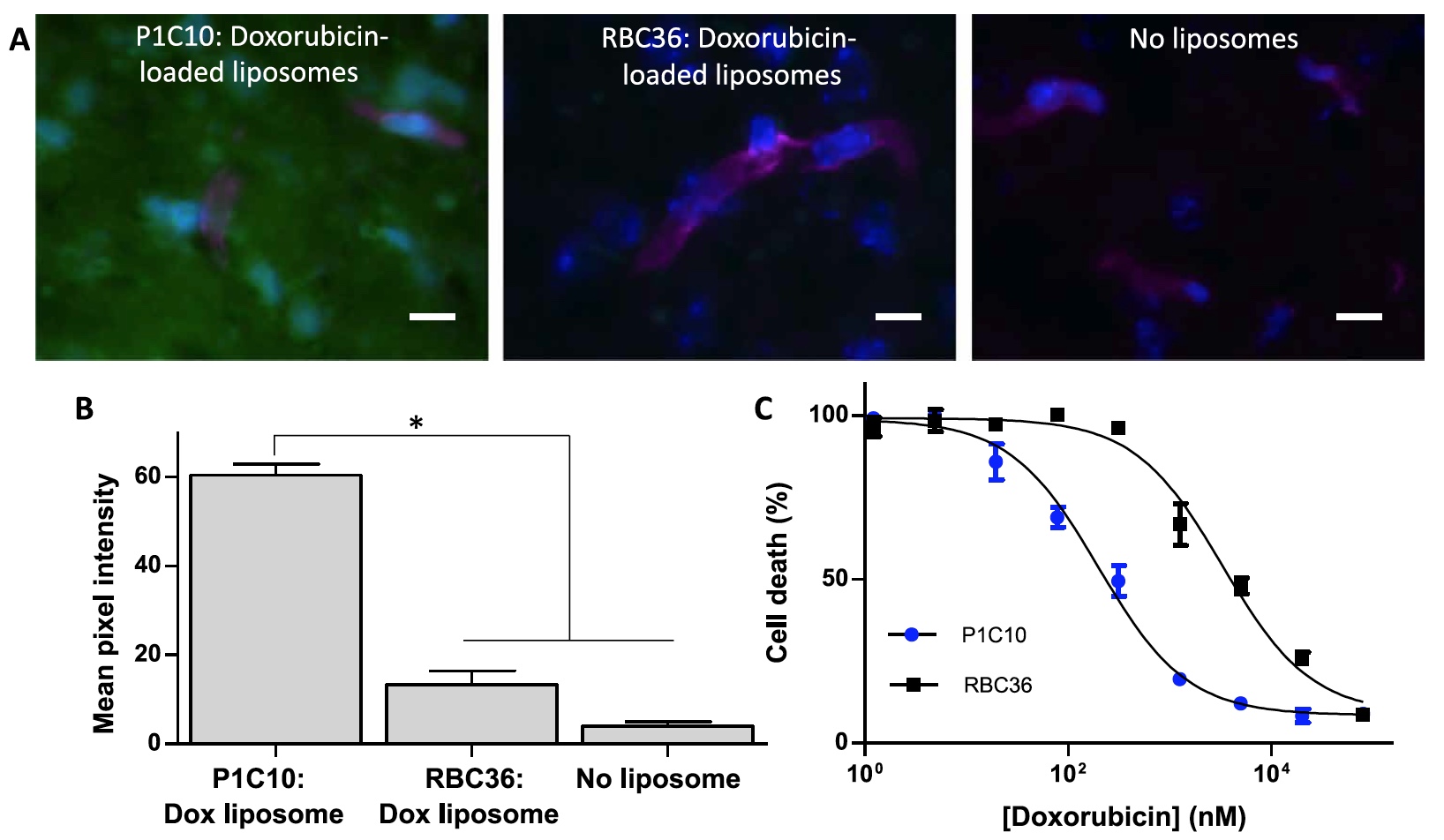
Image number 5
To demonstrate the possibility of treatment with doxorubicin-loaded liposomes targeting VLR, U87 tumor cells (cultured on bEnd.3 ECM) were incubated with doxorubicin-loaded liposomes targeting P1C10 or RBC36.
Observations showed a significant growth of the destroyed cells with liposomes targeted at P1C10. The half-maximal effective concentration was 199.0 ± 1.7 nM for P1C10 and 3312.0 ± - 2.6 for RBC36.
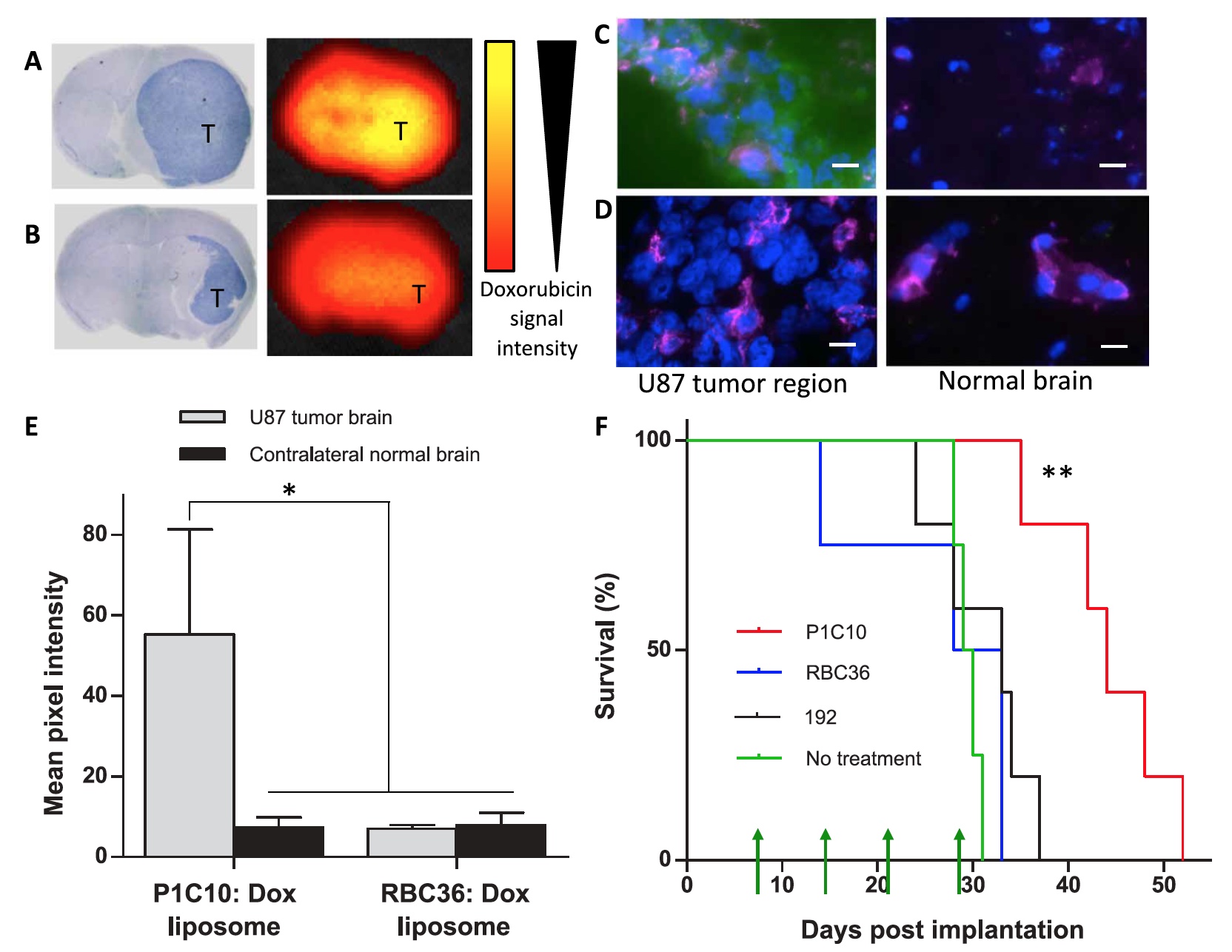
Image number 6
Finally, scientists tested the potential therapeutic utility of targeting the pathologically damaged extracellular matrix of the brain. For this purpose, doxorubicin-loaded liposomes were used in conjunction with P1C10, 192 or RBC36 in test mice with U87 cancer cells.
As can be seen in pictures 6a and 6b , the signal from doxorubicin is significantly stronger when using P1C10. At the same time, it is clear that this signal is sufficiently localized within the tumor and does not affect the contralateral (healthy) region of the brain.
Images 6c and 6d demonstrate that the accumulation of P1C10 in the tumor region is 7.6 times higher than in the healthy region. If we compare the degree of accumulation of P1C10 with RBC36, then the difference is 7.9 times in favor of P1C10.
For 4 weeks (7, 14, 21, and 28 days), 12 mg / kg of doxorubicin was administered to test mice with U87 using P1C10, 192, or RBC36.
After tumor implantation, the median survival was 43 days for P1C10, 30 days for 192 and 28 days for RBC36.
The group of experimental with P1C10 showed a significant increase in survival compared with the other two groups. At the same time, indicators 192 and RBC36 are not very different from those of the control groups that did not receive treatment at all.
For a more detailed acquaintance with the nuances of the study I recommend to look into the report of scientists .
Epilogue
There would be no happiness, but misfortune helped. This phrase can quite accurately describe this study. Scientists have applied pathological disorders of the hemato-encephalic barrier as a tool for drug delivery to certain areas of the brain affected by the tumor, while avoiding healthy areas. And scientists received help in this complex and noble undertaking, as they say, from where they did not expect. Variable lymphocyte receptors (VLR) lamprey became the basis of this work. Now you can say with confidence that even from parasites is good.
Treatment of brain tumors is associated with a huge list of difficulties, and the success of this process is not as great as we would like. Creating new methods and tools to combat such serious diseases is really what science is for. Knowing the world around and within ourselves, we do not go against the will of nature, we just begin to understand it better, we find new knowledge and apply it for the good.
Friday off-top:
“Desert Sea” (2011, voice overs - David Attenborough) - a documentary about two different neighboring underwater ecosystems.
Thank you for your attention, stay curious and have a great weekend, guys! :)
“Desert Sea” (2011, voice overs - David Attenborough) - a documentary about two different neighboring underwater ecosystems.
Thank you for your attention, stay curious and have a great weekend, guys! :)
Thank you for staying with us. Do you like our articles? Want to see more interesting materials? Support us by placing an order or recommending to friends, 30% discount for Habr's users on a unique analogue of the entry-level servers that we invented for you: The Truth About VPS (KVM) E5-2650 v4 (6 Cores) 10GB DDR4 240GB SSD 1Gbps $ 20 or how to share the server? (Options are available with RAID1 and RAID10, up to 24 cores and up to 40GB DDR4).
VPS (KVM) E5-2650 v4 (6 Cores) 10GB DDR4 240GB SSD 1Gbps before summer for free if you pay for a period of six months, you can order here .
Dell R730xd 2 times cheaper? Only we have 2 x Intel TetraDeca-Core Xeon 2x E5-2697v3 2.6GHz 14C 64GB DDR4 4x960GB SSD 1Gbps 100 TV from $ 199 in the Netherlands! Dell R420 - 2x E5-2430 2.2Ghz 6C 128GB DDR3 2x960GB SSD 1Gbps 100TB - from $ 99! Read about How to build an infrastructure building. class c using servers Dell R730xd E5-2650 v4 worth 9000 euros for a penny?
Source: https://habr.com/ru/post/453064/
All Articles