Hydrogel, blueberries and a pinch of turmeric: artificial vascular system

What is the most important organ in the human body? Romantics will say the heart, pragmatists will say the brain, and realists will say everything. And this is so, because the human body is a harmonious mechanism consisting of many parts, large and small, working in unison. If we talk about the most important fuel for such a mechanism, then one of the first in the head, of course, comes oxygen. And the oxygen supply is handled by the cardiovascular system. Today we will get acquainted with the study, in which scientists managed to create an artificial vascular labyrinth from a photopolymerizable hydrogel. How did they create artificial vessels, how effective are they, are they inferior in some real vessels, and what's the reason for turmeric? This and not only we will learn from the report of the research group. Go.
The basis of the study
The basis of artificial vessels, the main task of which is the transfer of fluid, is a material that works with liquids just fine. This material is called hydrogel.
')
A hydrogel is a collection of hydrophilic * polymer chains, sometimes found in the form of a colloidal gel in which water is a dispersion medium * .
Hydrophilicity * - the ability to absorb water well, the antipode of hydrophobicity (the ability of a molecule to repel water).
Disperse system * - a compound of several phases that do not mix and do not react chemically with each other. A prime example of a disperse system is air, cloud, composite materials, etc.A three-dimensional solid from a hydrogel is formed by cross-linking that holds hydrophilic polymer chains. Because of this, the structural integrity of the hydrogel network does not dissolve even with a high concentration of water. In this case, the hydrogel is an excellent absorbent.
Another important feature of this study for hydrogel is its flexibility, comparable to the flexibility of natural tissues, which is associated with a high water content.
Not only the material was unusual, but also the method of its application. Since the morphology of the vascular and pulmonary systems is very complicated and confusing, it would be wrong to use conventional 3D printing methods. Scientists have used stereolithography to create soft hydrogels containing the necessary vascular “labyrinths” inside.
Unlike standard extrusion printing, when voxels * are applied sequentially, photo stitching * allows you to use a projection image and create millions of voxels at the same time.
A voxel * is a volume image element, like a pixel in a two-dimensional image.
Photocrosslinking * (photocrosslinking) - photo-induced formation of a covalent bond between two macromolecules or between two different parts of the same macromolecule.In stereolithography, the resolution xy is determined by the passage of light, whereas the resolution z is determined by light-absorbing additives that absorb excess light and limit polymerization to the desired layer thickness, thereby improving the accuracy of the pattern of the object being created.
It should be clarified that the term “resolution” in three-dimensional printing has several definitions at once, due to the presence of three-dimensionality, that is, the x , y and z axes.If you do not use photo-absorbing additives, the hydrogel model will be extremely limited in terms of the shape and complexity of the structure. And the problem arises: to use conventional light-blocking chemicals that are used to structure photoresist or to manufacture plastic parts (for example, Sudan I - C 16 H 12 N 2 O) is impossible in bio-production because of the toxicity and carcinogenicity of such substances. But scientists are not so easily discouraged. They offered to use synthetic and natural food colors, which do an excellent job with photoabsorption and are safe for human health.
The xy resolution is the smallest movement made by a laser or extruder during the three-dimensional printing of one layer. The smaller this indicator, the more accurate the result will be. The resolution z is already the thickness of the layer itself.
Initially, the researchers tried to create a monolithic hydrogel consisting mainly of water and polyethylene glycol diacrylate with a cylindrical channel 1 mm in diameter oriented perpendicular to the axis of light projection. But even such a simple model is very difficult to create due to the fact that the low mass fraction of combinable elements and the need for longer polymerization lead to solidification in narrow channels, which must naturally be hollow.
In order to solve this problem, it was necessary to select certain constituent elements of the future model, including food dyes. Scientists have found that using tartrazine (yellow food dye, E102), curcumin (from turmeric) or anthocyanin (from blueberries) allows you to get a hydrogel with a vascular labyrinth without solidification, blocking the flow of fluid through the channel.
Among inorganic compounds, gold nanoparticles (50 nm) showed excellent results, which are distinguished by a high degree of light absorption and good biocompatibility.
Research results
Combining all the above-described discoveries and previous developments, the researchers proceeded to the practical implementation of a hydrogel containing a vascular grid.
First of all, tests of chaotic mixers (mixers), that is, intravascular topologies that homogenized * liquids as a result of interactions between fluid flows and vessel geometry, were carried out.
Homogenization * - the process of reducing the heterogeneity of the distribution of chemicals and phases in the volume of the total system for them.A monolithic hydrogel was created with a built-in static (fixed) mixer consisting of three-dimensional twisted blades (150 mm in thickness) with alternating chirality inside a 1-mm cylindrical channel.

Image number 1
To test the performance of such a mixer, laminar fluid flows were applied to a static mixer with a low Reynolds number (0.002). As a result, rapid mixing per unit length ( 1A ) was observed, depending on the number of blades.
Next, scientists created a three-dimensional bicuspid venous valve ( 1B ). The valves of this valve were dynamic (moving) and quickly responded to pulsating anterograde (forward movement) and retrograde (reverse movement) fluid flows. It is also worth noting the formation of stable vortices in the sinuses of the valve, which is fully consistent with the behavior of this valve.
Demonstration of the work of an artificial three-dimensional hydrogel bicuspid venous valve.
The next step is more complex and intricate vascular systems, which may consist of several labyrinths. The most important thing is that they should not intersect, otherwise there will be one big labyrinth as a result when two or more separate streams are necessary. Mathematical algorithms for filling space and fractal topology, applied by scientists, have shown good results in the design of two vascular labyrinths that do not intersect.

Image number 2
The researchers tested several versions of architectures with two non-intersecting channels: a spiral around a straight (axial) channel ( 2A ); Hilbert curves 1 ° and 2 ° ( 2B ); two-continuous cubic lattice ( 2C ); torus knot around torus ( 2D ).
Demonstration of all variants of vascular architecture, consisting of two independent channels.
Next, the scientists checked how effectively their artificial vascular system fulfills its main duties - the transport of oxygen. Liquid with deoxygenated erythrocytes (oxygen saturation ≤ 45%) passed through the spiral channel ( 2E ) enriched with moistened gaseous oxygen (7 kPa). At the exit, you can see the color change from dark red to bright red, which indicates the saturation of red blood cells with oxygen during the passage of fluid through the channel ( 2F and 2G ). Erythrocyte analysis after this test confirmed an increase in oxygen saturation.
Such a spiral vascular system is quite simple, as the scientists themselves say. And despite the excellent results in oxygenation, it is necessary to test the model under more severe conditions. The model of our lung is excellent for this, since in this case it is necessary to take into account not only the possibility of building a complex network of vessels, but also their elasticity - an important indicator in view of the dynamics of the lungs. Scientists, taking as a basis their previous developments and the works of their colleagues, created an alveolar model with an enveloping vascular network, which is based on the principle of the complex three-dimensional structure “Weir-Phelan foam”.

Image number 3
The Weir-Phelan foam is based on convex polyhedrons, but this does not prevent the creation of concave ones that will resemble alveolar air sacs with a common atrium of the airways ( 3A ). The resulting model consisted of 185 vascular segments and 113 intersection points.
Next, the model was applied for printing. The size of the bills was 5 pl, and the printing time was 1 hour ( 3B ). Cyclic ventilation of the combined respiratory tract with moistened gaseous oxygen led to a noticeable stretching and change in the curvature of the concave regions of the respiratory tract. Perfusion of deoxygenated erythrocytes at the entrance to the vascular system (from 10 to 100 mm / min) during cyclic ventilation led to a noticeable compression and clearance of erythrocytes from vessels adjacent to the concave regions of the respiratory tract ( 3C ).
Demonstration of the alveolar model with enveloping vascular mesh.
Computational model analysis data confirmed anisotropic stretching of the concave airway regions during inflation, i.e. expansion ( 3D ).
Given that the volume of hydrogel (0.8 ml) in the alveolar model is about 25% of the volume of the spiral model, the oxygenation efficiency of both models is almost identical ( 3E ).
Scientists believe that the branching (mesh) topology of a hydrogel and its stretching, as well as the redirection of flows during ventilation, allows increasing the absorption of oxygen by red blood cells, that is, their oxygenation.
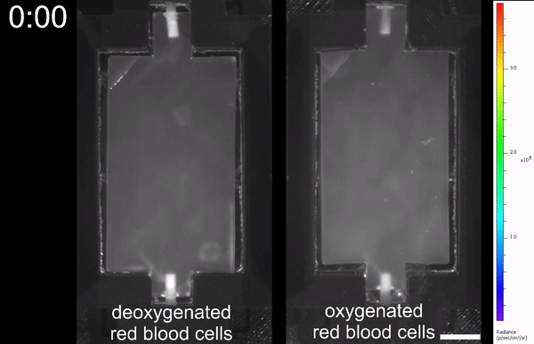
Comparison of deoxygenated (left) erythrocytes and oxygenated (right) erythrocytes inside the manufactured vascular system.
One of the most important moments is scalability. In other words, it is necessary to take into account the location of the entrance / exit of the vascular system and the air duct so that this architecture is as close as possible to the real lungs. The initial test volume of the hydrogel resulted in a very extensive system ( 3F ). Input and output vascular systems should be located at an angle of 180 degrees relative to each other and be topologically displaced from the respiratory tract. The vessels themselves must reach the farthest branches, that is, the alveolar vesicles consisting of 354 vascular segments and 233 vascular intersection points ( 3G ).
Testing of the obtained alveolar model showed that it is able to withstand more than 10,000 ventilation cycles at a pressure of 24 kPa and a frequency of 0.5 Hz for 6 hours. At the same time, during the test, both humidified oxygen and humidified nitrogen ( 3H , 3J ) were used.
The image 3I clearly shows that the developed system provides mixing of red blood cells and bidirectionality of flows within individual vessel segments.
Demonstration of a pulmonary model consisting of several alveolar.
The developed system shows excellent results during the tests, as we have already understood, but another important question remains - whether the hydrogel model is compatible with living cells.
In order to verify this, scientists used stereolithography to make the same models as described above, but already containing live mammalian cells. These cells were human mesenchymal stem cells. Analysis of the resulting system showed that the cells inside the hydrogel structure remain viable and can undergo osteogenic differentiation.
Such positive results could not be left without verification, because scientists decided to conduct a series of tests to establish the usefulness of this method of manufacturing biocompatible artificial systems.
The liver was taken for the basics, because this organ performs a number of important functions in the body, the success of which is strongly dependent on the structural topology of this organ.

Image number 4
The researchers created a complex hydrogel structure consisting of a variety of single-cell tissues and hydrogel carriers containing aggregates of hepatocytes ( 4A - 4C ).
The promoter activity of albumin tissue carriers containing aggregates was increased by more than 60 times compared with the activity of implanted tissues containing individual cells ( 4B , 4C ). In addition, upon careful examination of the tissues after resection, the hydrogel carrier tissue was more integrated with the tissue and blood of the test mouse ( 4D ).
Hepatic aggregates are better than individual cells, but they introduce difficulties in the process of creating hydrogel models, because their size exceeds the lowest resolution of voxels (50 mm).
In order to solve this problem, scientists have created their own architecture of carrier units ( 4E ). A network of microchannels was seeded with human umbilical vein endothelial cells as this improves tissue survival. Further, this artificial system was transplanted into the liver with chronic rodent injuries. 14 days after implantation, albumin promoter activity was observed, which indicates the survival of functional hepatocytes, that is, the viability of transplanted liver cells ( 4F ). Immunohistological analysis showed the presence of hepatic aggregates on the surface of the printed components of the hydrogel ( 4F and 4G ). In addition, the usual analysis of the images showed the presence of blood of the individual carrier within the implanted hydrogel system, which again confirms the absence of any rejection.
For more detailed acquaintance with the nuances and details of the study I recommend to look into the report of scientists and additional materials to it.
Epilogue
The result of this study is a vascular system based on hydrogel and natural / artificial food dyes, which copes well with its main tasks, in particular with the transport of oxygen. In addition, scientists used an off-the-shelf printing method (stereolithography), which allows you to create complex architectures in a relatively short time. In the future, scientists intend to improve their offspring, because the vascular system of each organ or body region has its own characteristics that need to be considered, studied and taken into account in the development of a more advanced hydrogel artificial counterpart.
The creation of artificial tissues, their aggregates and subsequently organs is a painstaking and very complex process. But good things are very often difficult. And this study can not be called anything other than a good deed. The first problem faced by a sick person in need of transplantation of an organ is waiting. For example, according to some sources in the US, 20 people die daily in a lineup for a liver transplant. The second problem is the donor. You can not just take the organ of one person and transplant it to another. Compatibility of a number of parameters is required. And the second problem smoothly feeds the first, extending the waiting time for the rescue operation.
Of course, mass cultivation of organs and systems, like tomatoes on a farm, with further transplantation is only the future for the time being, but how far it depends on such studies and their success. Speaking specifically about today's work, we can say that such a future has become a little closer.
Thank you for your attention, stay curious and have a good working week, guys!
Thank you for staying with us. Do you like our articles? Want to see more interesting materials? Support us by placing an order or recommending to friends, 30% discount for Habr's users on a unique analogue of the entry-level servers that we invented for you: The whole truth about VPS (KVM) E5-2650 v4 (6 Cores) 10GB DDR4 240GB SSD 1Gbps from $ 20 or how to share the server? (Options are available with RAID1 and RAID10, up to 24 cores and up to 40GB DDR4).
VPS (KVM) E5-2650 v4 (6 Cores) 10GB DDR4 240GB SSD 1Gbps before summer for free if you pay for a period of six months, you can order here .
Dell R730xd 2 times cheaper? Only we have 2 x Intel TetraDeca-Core Xeon 2x E5-2697v3 2.6GHz 14C 64GB DDR4 4x960GB SSD 1Gbps 100 TV from $ 199 in the Netherlands! Dell R420 - 2x E5-2430 2.2Ghz 6C 128GB DDR3 2x960GB SSD 1Gbps 100TB - from $ 99! Read about How to build an infrastructure building. class c using servers Dell R730xd E5-2650 v4 worth 9000 euros for a penny?
Source: https://habr.com/ru/post/450790/
All Articles