New technology can improve resolution of images of biological tissues tenfold.
The approach developed at MIT overcomes the long-standing problem of light scattering in biological tissues and other complex materials.
The problem of obtaining images of deep layers of biological tissues has long remained difficult. Light is usually scattered in such a complex material as biological tissue, and is reflected inside it until it comes back under many different angles. This disrupts the focusing of optical microscopes, reducing both the resolution and the depth of imaging. The use of light with an increased wavelength helps reduce scattering, but also reduces resolution.
Now, instead of trying to avoid scattering, MIT researchers have developed a technology that uses this phenomenon for their own purposes. The technology described by them in a paper published in the journal Science allows using light scattering to improve image resolution by a factor of 10 compared with existing systems.
The possibilities of conventional microscopes are limited by the diffraction limit , which does not allow focusing to be carried out with a more precise resolution. The new technology allows you to take pictures with "optical superresolution", overcoming this limitation.
')
It can be used to improve biomedical images, for example, concentrating more precisely on cancerous tissue cells. It can also be combined with optogenetic technologies to excite certain brain cells. It can even be used in quantum computing, as Dungu Kim, a graduate student, MIT mechanical engineer, lead author of the work, claims.
For the first time, researchers proposed this method in 2007 — by forming a light wave before sending it into a tissue in a special way, it is possible to reverse the scattering process and focus the light at one point. However, for a long time, it was not possible to take advantage of this method due to the complexity of collecting information on the scattering of light in such complex materials as biological tissues.
To obtain this information, researchers have developed various technologies to create "guiding stars", or feedback signals, emanating from certain points of the fabric, which allow the light to be correctly concentrated. But so far, these approaches have been giving permission that does not reach the diffraction limit, Kim says.
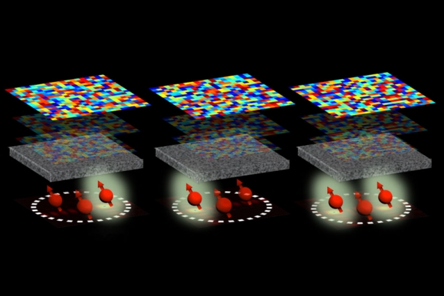
To improve resolution, Kim and co-author Dirk Inglund, an associate professor from the electrical engineering and computer science department of MIT and the electronics research lab, developed something called “reference quantum beacons” (QRM).
RCMs are created using nitrogen-substituted diamond vacancies (NV centers). These tiny molecular defects in the crystal lattice of diamonds have a natural fluorescence, that is, they emit light when excited by a laser beam.
Moreover, when a magnetic field is applied to the MCM, each of them resonates at its particular frequency. By sending a microwave signal of the same resonant frequency to a tissue sample as is observed at a certain RCM, researchers can selectively change its fluorescence.
“Imagine a pilot trying to navigate at night to a destination,” says Kim. “If he sees three beacons giving them a signal, he may be confused.” But if one of the lighthouses flashes on purpose, it will orient, ”he says.
In this sense, NV centers operate as beacons, emitting fluorescent light. By modulating the fluorescence of a particular beacon, researchers create an on / off signal and can determine the location of this beacon in tissue. “We can consider the location where the light comes from and, based on this, understand how light is scattered in complex materials,” says Kim.
The researchers then combine the information obtained from all the ACMs and create an accurate profile of the scattering pattern in the tissue. Using this picture with a spatial light modulator — a device used to obtain holograms through light manipulation — you can advance a certain shape to the laser beam to compensate for the scattering occurring inside the tissue. Then the laser will be able to focus with superresolution on a point inside the tissue.
Applied to the tasks of biology, the researchers suggest that weighted nanodiamonds can be introduced into the tissue, which will play the role of a contrast agent used in some methods of tissue imaging. Alternatively, molecular labels attached to diamond nanoparticles can deliver them to certain types of cells.
RCM can also be used as qubits for quantum sensors and quantum information processing, says Kim. “OKM can be used as quantum bits to store quantum information, so we can do quantum computing,” she says.
Obtaining images with superresolution in an environment with complex dispersion was previously hampered by the lack of “guiding stars” that would show their position with subdiffractional accuracy, as Vonshik Choi, a professor of physics from the Korean University who is not associated with this study, says.
“Researchers have developed an elegant method of operating RCM based on NV centers in nanodiamonds as such guiding stars,” he says. “This work opens up new possibilities for obtaining images of deep layers of tissues with superresolution and quantum information processing in nanodevices smaller than the wavelength”.
Now researchers hope to explore the possibility of using quantum entanglement and other types of semiconductors as an RCM, Kim says.
Source: https://habr.com/ru/post/442052/
All Articles