For the first time, a polymer prosthesis simulating bone structure has been successfully implanted
A team of scientists from a small innovative enterprise Biomimetix, which implements the development of NITU "MISiS", for the first time in the world successfully implanted a biomimetic hybrid prosthesis made from ultrahigh molecular weight polyethylene and titanium alloy into the femur bone simulating bone structure. The successful operation was carried out on the order of one of the Moscow veterinary clinics MedVet.
An implantation of an experimental biomimetic (similar in structure to the tissues of a living organism) prosthesis for a dog with an osteosarcoma of the femur was successfully performed in August 2018. After a few days, the animal was able to walk again, and, according to forecasts, mobility will soon recover completely.
Osteosarcoma (osteogenic sarcoma) is a fast-growing cancer, whose cells are derived from bone tissue, lead to its gradual destruction and, accordingly, loss of mobility. Osteosarcoma is the most common type of bone tumor in both humans and animals.
Treatment necessarily includes a course of chemotherapy and surgery with removal of the affected tissue. Advanced technologies allow organ-preserving surgery - in this case, instead of a remote part of the bone, a metal, ceramic or polymer implant is placed. Despite the fact that such prostheses allow to restore mobility, they differ greatly in structure from bone tissue, and this can lead to a number of significant difficulties.
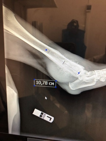
Fig. 1 - X-ray of the hind limb of the animal before the operation. Osteosarcoma affects almost 11 cm of bone tissue.
“Traditional materials for medical prosthetics have a number of significant drawbacks: for example, titanium implants take on too much pressure for bone, and the latter begins to thin. In such a situation, the bone at the junction with the prosthesis may break. Another option is ceramics, but it is more fragile, which may limit the size of recoverable bone tissue. In addition, the structure of these materials does not allow them to “grow together” with the bone - constant tight fixation is required, ”explains Fedor Senatov, General Director of Biomimetix, research associate at the Center for Composite Materials, NITU“ MISiS ”.
The Biomimetix team has been developing biomimetic implants for several years. Scientists work with ultra-high molecular weight polyethylene - a proven biocompatible material - but modify it in a unique way, giving implants the desired structure. This is the first experience in the world of creating biomimetic prostheses from such a polymeric material.
')

Fig. 2 - X-ray of the hind limb of the animal after implant placement.
In August of this year, by order of the MedVet veterinary centers network, the Biomimetix team developed an experimental biomimetic prosthesis of the femur for a dog with osteosarcoma.
“Since the dog turned out to be large - a dog, she was accustomed to move actively, and it took about 11 cm of bone to be removed, it was decided to make the prosthesis hybrid. On a titanium tube made by 3D printing by our partners, Konmet, we expanded a layer of continuous ultrahigh molecular weight polyethylene, and the inner part was made of porous ultra high molecular weight polyethylene, identical to the structure of the cancellous bone. During the operation, part of the cover was cut off in order to “fit” the implant to the bone. Within a few days after the operation, the dog was able to walk fully. If the polymer grows together with the bone tissue successfully, after some time it will be possible to remove the fixing plates, ”says Fedor of the Senates.

Fig. 3 - Implants with a titanium reinforcing component, "spongy" polymer core and "cortical" polymer sheath.
“My colleagues and I successfully performed a prosthesis operation. The implant is durable, its size corresponds to the weight of the dog and the size of the bone. Here, long-term results are more important, so that the implant gets used, grows into the bone. Then there will be a lot of progress, but it takes time. While we are watching a dog, she is now undergoing chemotherapy, ”says the head physician of MedVet, Ph.D. Ilya Vilkovysky.
According to the developers, the preparation took 2 weeks, during which time they did:
- development of implant design by X-ray,
- 3D modeling,
- carrying out computer simulations,
- development of engineering solutions to change the technology of creating a hybrid implant,
- obtaining test samples
- carrying out mechanical tests,
- making the final implant.
Source: https://habr.com/ru/post/425751/
All Articles