MIS. Inserts and removed organs
In the IIA, the research protocol pattern is more like a constructor, which may consist of parts of different shapes and sizes. As a building material are the previously discussed tags and their attributes . With their help, you can add to the protocol all the fields that must be in it. However, sometimes there are cases when it is necessary to expand the capabilities of the current study protocol by adding additional measurement units to it. We called such additional parts inserts. In general, inserts can be an unlimited number. Templates of additional parts consist of the same tags as the template of the protocol itself, but other tags are used to indicate the insertion point and description of its type.
The description of the distant organs or anatomical parts of the body also requires special attention. They have nothing to do with the inserts, since the potential organs for removal are strictly in the standard study protocol. By default, the template contains all the anatomy described, and the absence is regulated by a special tag. It contains the status of the corresponding anatomy.
')
Let us consider in more detail the mechanics of the work of additional tags.
Inserts
One of the types of inserts - education . Its characteristic features are:
- the described object is initially absent in a healthy organism and appears over time due to some circumstances;
- the same type of the described object may be present in the body in more than one quantity.
Using the insert with the type of "education", you can describe the following types of pathologies:
- some pathological processes - a sequence of reactions that occurs due to the action of a pathogenic factor - a neoplasm (node, cyst, tumor), atheromatous plaques (contribute to the development of atherosclerosis), and so on;
- protrusions and hernias;
- stones (stones);
- wounds, etc.
If a doctor can familiarize himself with the dynamics of changes and comments for “standard” organs, then for formations in addition to the types of dynamics already mentioned, the dynamics of the development of education itself is also important. For this was added another entity - anatomy addon . In addition to viewing the dynamics, the preservation of each formation contributes to the description of the existing formations during the next visits of the patient, and not to duplicate the same ones.
Taking into account the fact that one type of described pathologies may occupy different organs or one organ in different places, such a concept as localization was introduced. Firstly, it allows you to clearly understand where the pathology is located or where exactly the measurement was taken. Secondly, it becomes possible to decompose the pathology only once in the “anatomy” and “measurement” entities, and not to create this pathology several times for each organ.
Consider a cyst. It is a pathological cavity in tissues or organs that has a wall and contains fluid. Its size, structure and content depends on many factors, and localization is one of them. According to the concentration of cysts within the same localization single and multiple cysts are isolated. Instead of creating anatomy of a single cyst of the left thyroid lobe, a single cyst of the right thyroid lobe, a single pancreatic cyst, etc., a single anatomy of a single cyst with the necessary measurements was added to the database. In turn, anatomy is already a localization: the left lobe of the thyroid gland, the right lobe of the thyroid gland, the pancreas, etc.
There is one anatomy in the human body, the status of which is not abated by disputes in various fields of science to this day. This is the fruit. For example, lawyers argue about the rights of an embryo, in bioethics they raise the question of recognition of the status of a person as the embryo and a fetus, etc. Within the framework of the IIA, we identified two important features for the anatomy of the fetus. First, this anatomy appears in the female body, and is not present there originally. Secondly, several fetuses can develop at once in the womb (multiple pregnancy). As the saying goes: “if something looks like a duck, swims like a duck and quacks like a duck, then this is probably what a duck is.” Taking into account the characteristic features of this anatomy, and not its essence, the fetus is added to the study protocol as the pathologies discussed above - with the help of the insert “education”.
The second type of inserts - additionally described anatomy , which we abbreviate as - additional anatomy . In contrast to the objects described through the insert "education", the insert "additional anatomy" is intended for anatomy that are present in the body initially, but if they are not in an altered state, then they are not described. If the anatomy is changed, then it falls into the anatomy addon . Such anatomies include: the vertebrae of all parts of the spine, parathyroid gland, sublingual salivary glands, mammary glands, etc.
The third type of inserts is the extra dimensions . A few examples:
- in the study protocol of the examination by a urologist, measurements are added depending on the gender of the patient;
- in the protocol of the pediatrician's study, the inserts measure for children under one year old and the reflexes of children under one year old;
- a positive test for hepatitis C in the study "Anti-HCV antibodies (Anti-HCV)" test is performed to confirm the diagnosis of "viral hepatitis C", which checks for the presence of antibodies to some hepatitis C proteins;
- additional tests for the presence of salts and bacteria can be made in the analysis of urine;
- bacteriological sowing - microbiological laboratory testing of biological material by sowing it on certain nutrient media in order to detect the presence of pathogenic and conditionally pathogenic microorganisms in it - when confirming any bacteria using additional measurements, the sensitivity of this microorganism to a specific preparation is specified.
The inserts themselves are indicated in the template by the tags insertion-place and insertion-template-builder . A special button appears in the editable conclusion form on the place of the insertion-place tag. When you click on it, the doctor displays a dialogue with all possible formations, additional anatomy and additional measurements that are indicated in the template of this study protocol.
Insertion place
Used to indicate where to insert. Contains the insertion-template-builder tag.
Insertion-template-builder
Used to indicate a specific formation, additional anatomy or additional dimensions:
- id - number of the template in the database. Multiple values may be specified;
- anatomy-id - the number of anatomy in the database. Indicated for formations and additional anatomy;
- localization-id - the number of anatomy for localization in the database. Multiple values may be specified. Indicated for formations;
- name-overriden is the name of the insert. For formations and additional anatomy, the name of the anatomy is taken from the database by default;
- name-overriden-button - the names of the buttons in the insert selection dialog. The default value is from name-overriden;
- use-template-builder-name - flag to display the name of the template specified in the database. Options: true and false. Default is false;
- localization-marker-type - marker for the localization field. Options: none, word (spelled "Localization:"), icon (icon is inserted). The default is none;
- need-anatomy - a flag to display the anatomy field. Options: true and false. The default is true;
- need-name - flag to display an additional name field. Options: true and false. The default is true;
- need-status - flag to display the status field. Options: true and false. The default is true;
- need-localization - flag to display a drop-down list with localizations. Options: true and false. The default is true;
- need-found-date - flag to display the date field. Options: true and false. The default is true;
- need-template-builder — flag to display a drop-down list with template views. Options: true and false. The default is true;
- anatomy-width, name-width, status-width, localization-width, found-date-width, template-builder-width — adjust the width of the anatomy, name, status, localization, date, name of the template, respectively;
- font-bold, font-underline - font settings;
- spacing-before, is-short .
Insertion picture
Sometimes there are complex localizations for which a verbal description (a clear indication of anatomy-localization) may not be enough. For such cases, a special component was developed. It is a picture-substrate (anatomy-localization image) with areas that you can click on. The selected area (location of the formation) is highlighted in the specified color.
- id - number of complex localization in the database;
- selection-color - the highlight color of the selected area in the image-substrate. The default is # 000000 (black);
- spacing-before, is-short .
This component can be used to indicate protrusions / hernias of intervertebral discs, atheromatous plaques in vessels, etc.

Of education
Let's go back to the cysts. They can be formed due to disorders arising in the period of embryonic development, traumas, infections, genetic predisposition, inflammatory processes, stress, etc. This neoplasm may change in size over time. In some cases, it resolves itself. When a cyst reaches a certain size, it begins to interfere with nearby organs, causing unpleasant sensations in a person.
Some cysts can be detected independently. For example, on the chest, skin, retention lip cysts. The latter appear on the surface of the tongue, cheek, lower lip. They are usually caused by biting or other injuries. Cysts in the internal organs are diagnosed using various methods of examination (ultrasound, MRI, etc.).
Consider the addition of single and multiple cysts to the protocol for an ultrasound examination of the thyroid gland.
Single cyst pattern:
<template> <line> <measurement id="2401" max-width="246" comment=""/> <measurement id="2402" measurement-name="" comment=" "/> <measurement id="2421" max-width="51" empty-name="true" comment=" "/> </line> <measurement-group id="83" is-short="false"/> <anatomy-comment comment-id="302" comment-type="comment" spacing-before="NONE" use-default="false" comment=" " /> </template> Multiple cyst pattern:
<template> <line> <measurement id="2423" max-width="246" comment=" "/> <measurement id="2424" measurement-name=" " comment=" "/> <measurement id="2425" max-width="51" empty-name="true" comment=" "/> </line> <measurement-group id="84" is-short="false"/> <anatomy-comment comment-id="303" comment-type="comment" spacing-before="NONE" use-default="false" comment=" "/> </template> Part of the thyroid pattern for adding a cyst:
<insertion-place> <!-- --> <insertion-template-builder anatomy-id="821" anatomy-width="100" name-width="136" need-template-builder="false" localization-id="23,1021,1022,1023,24,1024,1025,1026,25" need-status="false" localization-width="177" localization-marker-type="word" id="538"/> <!-- --> <insertion-template-builder anatomy-id="822" anatomy-width="100" name-width="136" need-template-builder="false" localization-id="23,1021,1022,1023,24,1024,1025,1026,25" need-status="false" localization-width="177" localization-marker-type="word" id="540"/> </insertion-place> Anatomy for localization (localization-id)
- 23 - the left lobe;
- 1021 - the lower third of the left lobe;
- 1022 - the middle third of the left lobe;
- 1023 - the upper third of the left lobe;
- 24 - right lobe;
- 1024 - the lower third of the right lobe;
- 1025 - the middle third of the right lobe;
- 1026 - the upper third of the right lobe;
- 25 - isthmus
Insert dialog:
When single and multiple cysts are added to the study protocol, we get the following result:

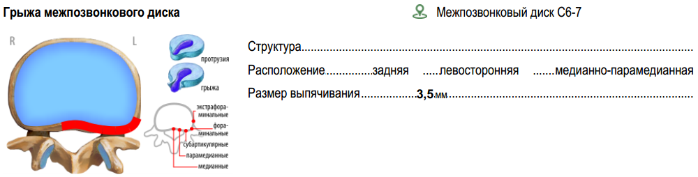
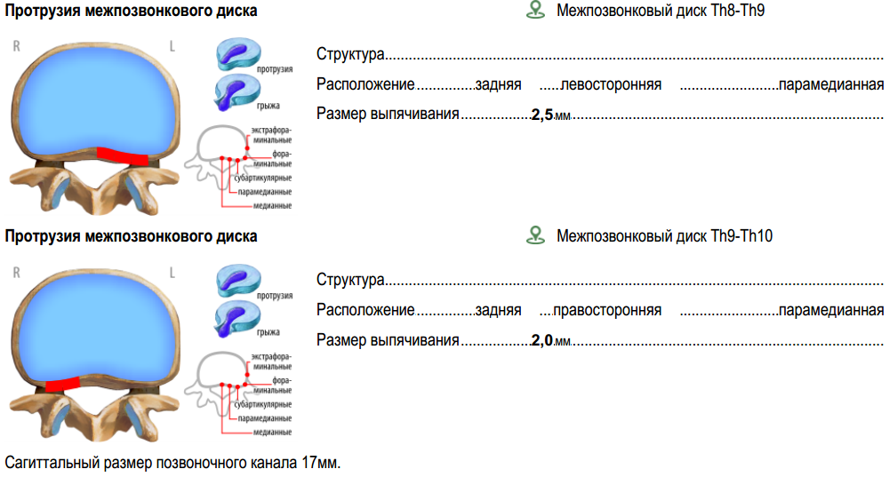
A sedentary lifestyle, excess weight, curvature of the spine, weak back muscles, osteochondrosis, serious loads on the spine, and a metabolic disorder - this is what causes the appearance of changes in the intervertebral discs. The intervertebral disk receives all the nutrients from neighboring tissues through diffusion, since there are no blood vessels in the disk itself. It consists of the pulpal (gelatinous) nucleus and the fibrous ring surrounding it. When the metabolism stops, dystrophic changes begin in the fibrous ring, which leads to its thinning, loss of elasticity and the appearance of cracks, and the pulpous nucleus begins to slowly move to the side. When the gelatinous core reaches the edge of the disk, it begins to go beyond its limits, stretching the fibers of the annulus. So protrusion is formed. If the fibrous ring is broken, then the protrusion begins to be considered a hernia.
For the description of protrusions and hernias complex localization is used. The pattern structure for them is the same:
<template> <insertion-picture id="3" is-short="true" selection-color="#FFB6AC" /> <measurement id="2622" is-short="true" comment=""/> <line is-short="true"> <measurement id="8821" measurement-name="" comment=" \"/> <measurement id="8822" max-width="70" empty-name="true" comment=" "/> <measurement id="8823" empty-name="true" comment=" "/> </line> <measurement id="2624" is-short="true" comment=" "/> <anatomy-comment comment-id="279" comment-type="comment" spacing-before="NONE"/> </template> Part of the MRI template of the thoracic spine:
<insertion-place> <insertion-template-builder anatomy-width="145" name-width="28" status-width="30" localization-width="150" found-date-width="55" template-builder-width="45" anatomy-id="901" localization-id="869, 1501, 1502, 1503, 1504, 1505, 1506, 1507, 1508, 1509, 1510, 1511, 1450" id="582,542" use-template-builder-name="true" localization-marker-type="icon"/> </insertion-place> In the id attribute, the value of 582 is the number of the protrusion pattern, and 542 is the hernia. Anatomy 901 ( anatomy-id attribute) - herniated intervertebral disc.
Anatomy used for localization (localization-id)
- 869 - intervertebral disk C7-Th1;
- 1501 - intervertebral disk Th1-Th2;
- 1502 - intervertebral disk Th2-Th3;
- 1503 - intervertebral disk Th3-Th4;
- 1504 - intervertebral disk Th4-Th5;
- 1505 - intervertebral disk Th5-Th6;
- 1506 - intervertebral disk Th6-Th7;
- 1507 - intervertebral disk Th7-Th8;
- 1508 - intervertebral disk Th8-Th9;
- 1509 - intervertebral disk Th9-Th10;
- 1510 - intervertebral disk Th10-Th11;
- 1511 - intervertebral disk Th11-Th12;
- 1450 - intervertebral disk Th12-L1.
Additional anatomy
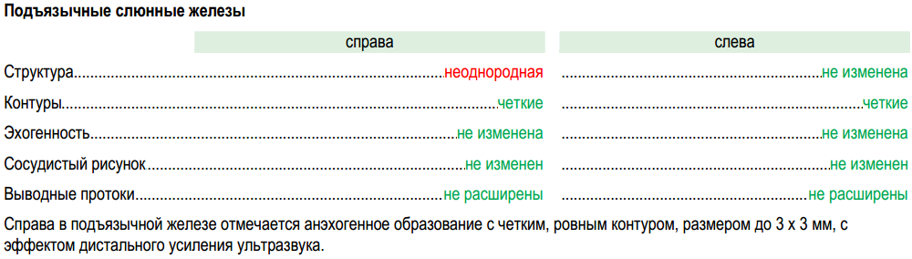
Consider the ultrasound pattern "soft tissues and lymph nodes of the neck, salivary glands", where you can describe the hyoid salivary gland.
Template sublingual salivary gland
<template> <line is-short="false" comment="-"> <text text-label=" " /> <text text-label="" max-width="193" is-color-selection="true"/> <text text-label="" max-width="193" is-color-selection="true"/> </line> <line is-short="false"> <measurement id="3969" comment=" " /> <measurement id="3981" max-width="193" empty-name="true" comment="" /> </line> <line is-short="false"> <measurement id="3970" comment=" " /> <measurement id="3982" max-width="193" empty-name="true" comment="" /> </line> <line is-short="false"> <measurement id="3971" comment=" " /> <measurement id="3983" max-width="193" empty-name="true" comment="" /> </line> <line is-short="false"> <measurement id="3972" comment=" " /> <measurement id="3984" max-width="193" empty-name="true" comment="" /> </line> <line is-short="false"> <measurement id="3973" comment=" " /> <measurement id="3985" max-width="193" empty-name="true" comment="" /> </line> <line is-short="false"> <measurement id="12481" comment=" " /> <measurement id="12501" max-width="193" empty-name="true" comment="" /> </line> <anatomy-comment comment-id="379" is-conclusion="false" spacing-before="NONE" comment=" " /> </template> Main pattern
<template> <line> <anatomy id="1261" font-size="10" font-bold="true" font-underline="false" is-short="false" comment=" " /> <measurement id="3861" comment=" " /> </line> <line is-short="false" comment="-"> <text text-label=" " /> <text text-label="" max-width="193" is-color-selection="true"/> <text text-label="" max-width="193" is-color-selection="true"/> </line> <template-builder id="597" comment=" "/> <template-builder id="598" comment=" "/> <insertion-place> <insertion-template-builder anatomy-width="180" need-template-builder="false" anatomy-id="1283" id="599" comment=" "/> <insertion-template-builder anatomy-width="180" need-template-builder="false" anatomy-id="1307" id="595 comment=" "/> </insertion-place> <anatomy-comment comment-id="111"/> <conclusion-label spacing-before="HALF"/> <anatomy-comment comment-id="51" comment-type="conclusion"/> </template> 
Additional measurements
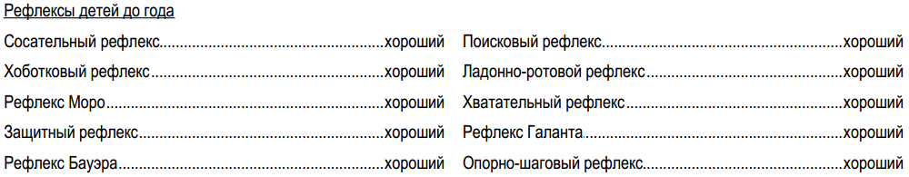
The previously considered Bauer reflex is in the insert with the reflexes of children up to one year old.
Insert Template:
<template> <line> <measurement id="8621" comment=" " /> <measurement id="8622" comment=" " /> </line> <line> <measurement id="8623" comment=" " /> <measurement id="8624" comment="- " /> </line> <line> <measurement id="8625" comment=" " /> <measurement id="8626" comment=" " /> </line> <line> <measurement id="8627" comment=" " /> <measurement id="8628" comment=" " /> </line> <line> <measurement id="8629" comment=" " /> <measurement id="8630" comment="- " /> </line> </template> Part of the pediatrician examination template:
<insertion-place> <!-- --> <insertion-template-builder font-bold="false" font-underline="true" need-template-builder="false" name-overriden-button=" " name-overriden=" " anatomy-width="200" id="921"/> <!-- --> <insertion-template-builder font-bold="false" font-underline="true" need-template-builder="false" name-overriden-button=" " name-overriden=" " anatomy-width="200" id="922"/> </insertion-place> 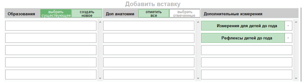
We will deal with the addition of a confirmatory test for hepatitis C. The test pattern itself:
<template> <measurement id="2365" comment=" (Anti HCV)"/> <measurement id="2362" comment=" Core (Anti HCV-Core)"/> <measurement id="2381" comment=" NS3 (Anti HCV-NS3)"/> <measurement id="2363" comment=" NS4 (Anti HCV-NS4)"/> <measurement id="2364" comment=" NS5 (Anti HCV-NS5)"/> </template> Template "Anti-HCV Antibodies (Anti HCV)":
<template> <measurement id="90" comment=" (Anti HCV) IgG"/> <insertion-place> <insertion-template-builder font-bold="false" font-underline="true" need-template-builder="false" anatomy-width="180" name-overriden=" . " id="572"/> </insertion-place> <anatomy-comment comment-id="355" comment-type="comment" spacing-before="NONE" comment=". "/> </template> 

Description of the removed organs
As you know, a person can adapt to life without certain organs. However, history remembers times when wild thoughts about “extra organs” appeared in the minds of scientists, which do not affect the vital activity of the organism and are subject to removal, and the sad consequences of their realization in reality. So in the late XIX - early XX century, this idea reached its apogee.
More about dashing times
Charles Darwin in his book "The Origin of Man and Sexual Selection" (1871) listed a number of rudimentary organs (appendix, bacterial bone, wisdom teeth, ear muscles, body hair, etc.), the presence of which, in his opinion, confirmed the theory of evolution.
The German anatomist Robert Widersheim in his work “The Structure of a Man from a Comparative-Anatomical Point of View” (1890) gave an extensive list of rudimentary organs. To the previously reviewed Darwin added tonsils, thyroid gland, tear glands, some vein valves, etc.
The Russian biologist Ilya Ilyich Mechnikov in his work “Studies on Human Nature” (1913) mentions the works of Darwin and Wiedersheim, and also discusses the imperfection and disharmony of the human body. One of the chapters is devoted to the digestive system, where the uselessness of the appendix is mentioned, and the suggestion made by the Dutch physician Kolbrugg about the usefulness of the appendix (appendix), as it develops intestinal bacteria that protect the body from certain diseases of the digestive system, is strongly rejected. However, Kolbrugg did not present any weighty evidence in favor of his theory. Mechnikov fell into disgrace and colon, the useful functions of which were minimized, but the problems associated with it, on the contrary, were considered in great detail (constipation, absorption of harmful substances, a focus of dangerous diseases and the frequent location of malignant tumors). Therefore, in the opinion of Mechnikov, the removal of this body as a whole would lead to positive results. Although the scientist himself did not check his statement, it was confirmed in the practice of some surgeons who performed such operations. There were also doctors who reacted to his words with great enthusiasm and sometimes cut off the organ unnecessarily. However, not all patients survived or could continue their normal life. Only after decades this method of treatment was seriously criticized.
Many doctors liked the idea of removing the appendix in advance, as many patients died of appendicitis. Do not hesitate to remove and tonsils with glands. The idea of removing these organs in children at an early age was most common in Europe and the USA. However, some time later, experts recognized their mistakes. Children without an appendix poorly absorbed breast milk, were lagging behind in development, were more often ill and had problems with digestion. The doctors did not understand why this was happening. Only in 2007, American scientists from Duke University (Duke University) found out that the appendix contains bacteria necessary for the normal functioning of the intestine. The role of the tonsils and glands was also learned later, and until that moment, doctors noticed that children without them are more likely to suffer from diseases of the bronchi and lungs.
In general, the development of the idea of "extra" organs can be explained by a banal ignorance about the work and functions of the alleged rudiments.
Sometimes practiced rather strange methods of treating certain diseases. Some of them were based on the removal of organs. For example:
- treatment of female hysteria, which began in the Victorian era and ended in Europe at the end of the XIX century, and in America - at the beginning of the XX century. Female genitals were subject to removal;
- treatment of mental illness by Henry Cotton. The doctor believed that such diseases cause infections. First, he removed the teeth of patients. If there was no effect, then the rest of the organs (glands, gallbladder, etc.) were used.
Today, doctors use the removal of organs and anatomical parts of the body (ectomy) only in extreme cases: injuries or serious pathologies. It can be removed as one of the paired organs (eyes, kidney, lung, etc.) in the case of normal operation of the other, or unpaired. The latter include:
- stomach - in the treatment of gastric cancer. After gastrectomy, the esophagus and small intestine are connected;
- spleen - in case of blood diseases, injuries and other pathological conditions;
- gallbladder - with cholecystitis;
- part of the liver - can be removed up to 75%. The liver has a regenerative function, which occurs very slowly;
- pancreas;
- colon - with Crohn's disease and malignant tumors;
- thyroid;
- etc.
An anatomy-status tag has been added to capture information about a remote organ or anatomical part of the body.
Anatomy-status
It is a drop-down list of three statuses: yes, no, not visualized. To correctly display the study protocol, it is necessary to take into account the peculiarities of the language. So for each gender and number, you can adjust the status options separately, and for anatomy - the desired list. For example:
- feminine, singular - is (visualized as a space in the document), deleted, not rendered;
- masculine unit number - “space”, deleted, not rendered;
- the plural is “space”, deleted, not rendered.
In addition to the obvious statuses, there is / no "not rendered" was added. As the name implies, it is used in cases where a body is not visible during the examination. For example, due to the accumulation of gas in the intestine, some abdominal organs may not be visible.
In general, an entity with anatomy statuses is used not only for the anatomy-status tag and organs to be removed, but also in inserts with the type “education”. For example:
- for the fetus - visualized, born, not visualized;
- for formations (cyst, node, etc.) - visualized, promptly deleted, not visualized.
The anatomy-status tag works as follows: if you select the “no” and “not visualized” statuses, all dimensions related to this anatomy are removed, and a line with the name of the anatomy and its status remains. In order for this to work, all the code related to the anatomy to be removed is separated into a separate template and a link to it ( template-builder tag) in the main template is written.
When the status of anatomy is set to "no" or "not visualized," it is stored in anatomy-addon .
Thus, the following anatomy can fall into the essence of anatomy-addon :
- changed anatomy, which usually do not describe;
- remote anatomy;
- appeared "non-standard" anatomy.
The anatomy-status tag contains several attributes:
- anatomy-id - the number of anatomy in the database;
- max-width - the overall width of the element;
- spacing-before, is-short .
The thyroid ultrasound pattern is one of those patterns that have undergone significant structural changes during system design.
The very first version of the thyroid ultrasound template
<template image-id="5" need-warning="true" image-position="left-top-corner"> <anatomy id="22" font-size="10" font-bold="true" font-underline="false" is-short="true" comment=" " /> <line is-short="true" spacing-before="HALF"> <measurement id="310" max-width="156" comment=""/> <measurement id="341" max-width="156" comment=""/> </line> <line is-short="true"> <measurement id="308" max-width="156" comment=""/> <measurement id="342" max-width="156" comment=""/> </line> <anatomy id="24" font-size="10" font-bold="false" font-underline="false" anatomy-name=" :" is-short="true" spacing-before="HALF"/> <measurement-group id="2" is-short="true" is-color-selection="true"/> <anatomy id="23" font-size="10" font-bold="false" font-underline="false" anatomy-name=" :" is-short="true" spacing-before="HALF"/> <measurement-group id="1" is-short="true" is-color-selection="true"/> <measurement id="307" is-short="true" measurement-name=" "spacing-before="HALF"/> <measurement id="309" measurement-name=" "/> <anatomy-comment comment-id="9" is-conclusion="false" spacing-before="HALF"/> <anatomy-comment comment-id="8" is-conclusion="true" spacing-before="HALF"/> </template> Adding template-builder and anatomy-status tags and inserts
Main template:
Right share:
Left lobe:
<template image-id="5" need-warning="true" image-position="left-top-corner"> <anatomy id="22" font-size="10" font-bold="true" font-underline="false" is-short="true" comment=" "/> <line is-short="true" spacing-before="HALF"> <measurement id="310" max-width="156" comment=""/> <measurement id="341" max-width="156" comment=""/> </line> <line is-short="true"> <measurement id="308" max-width="156" comment=""/> <measurement id="342" max-width="156" comment=""/> </line> <template-builder id="253" is-short="true"/> <template-builder id="254" is-short="true"/> <measurement id="307" is-short="true" measurement-name=" " spacing-before="HALF" /> <measurement id="309" measurement-name=" "/> <insertion-place> <!-- --> <insertion-template-builder anatomy-id="1041" anatomy-width="100" name-width="136" need-template-builder="false" localization-id="23,1021,1022,1023,24,1024,1025,1026,25" need-status="false" localization-width="177" localization-marker-type="word" id="578"/> <insertion-template-builder anatomy-id="1042" anatomy-width="100" name-width="136" need-template-builder="false" localization-id="23,1021,1022,1023,24,1024,1025,1026,25" need-status="false" localization-width="177" localization-marker-type="word" id="580"/> <!-- --> <insertion-template-builder anatomy-id="821" anatomy-width="100" name-width="136" need-template-builder="false" localization-id="23,1021,1022,1023,24,1024,1025,1026,25" need-status="false" localization-width="177" localization-marker-type="word" id="538"/> <insertion-template-builder anatomy-id="822" anatomy-width="100" name-width="136" need-template-builder="false" localization-id="23,1021,1022,1023,24,1024,1025,1026,25" need-status="false" localization-width="177" localization-marker-type="word" id="540"/> <!-- --> <insertion-template-builder font-bold="false" font-underline="true" need-template-builder="false" anatomy-width="180" name-overriden=" " id="537"/> </insertion-place> <anatomy-comment comment-id="9" comment-type="comment" spacing-before="HALF"/> <conclusion-label spacing-before="HALF"/> <anatomy-comment comment-id="8" comment-type="conclusion"/> </template> Right share:
<template> <line is-short="true"> <anatomy id="24" font-size="10" font-bold="true" font-underline="false" spacing-before="HALF" comment=" "/> <anatomy-status anatomy-id="24" /> </line> <measurement-group id="2" is-color-selection="true" is-short="true"/> </template> Left lobe:
<template> <line is-short="true"> <anatomy id="23" font-size="10" font-bold="true" font-underline="false" spacing-before="HALF" comment=" "/> <anatomy-status anatomy-id="23" /> </line> <measurement-group id="1" is-color-selection="true" is-short="true"/> </template> 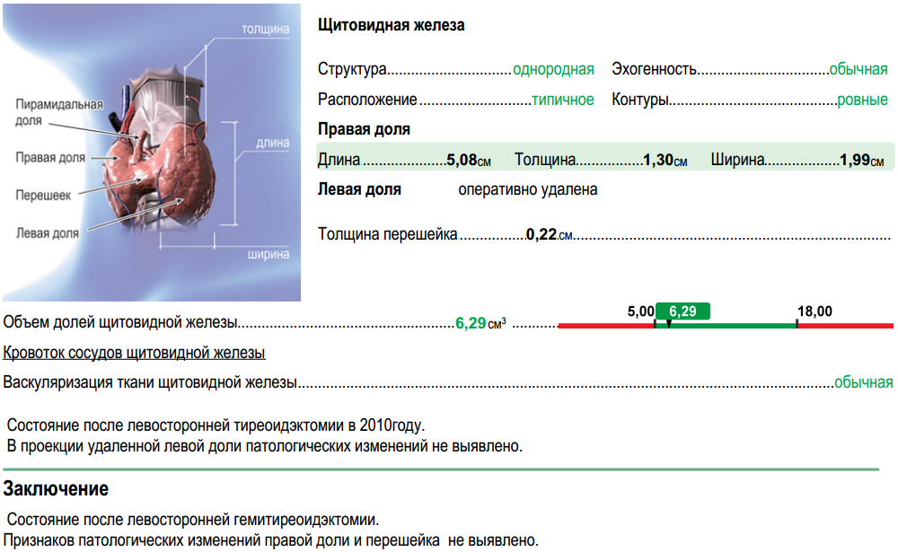
When we started, each research protocol template consisted of a comment and a conclusion. The desire for formalization resulted primarily in the creation of a system for storing numerical data and a description of human anatomy. As a result, comments declined and were used as explanations of the main data.
If you look at the conclusions, then among the information that usually gets there, we can distinguish the following types of data:
- diagnoses:
- "Signs of chronic tonsillitis"
- "duodenitis?"
- "K21.0 GERD with esophagitis"
- destination:
- "Recommended diet with restriction of fat"
- "Chlorhexidine 2-3 jets 2 times a day"
- directions:
- "general urine analysis"
- "MRI of the cervical spine"
- "Consultation endocrinologist"
Thus, the conclusion remains the "den" of non-formalized information. Therefore, the further development of the medical data storage system is aimed at formalizing these entities.
Source: https://habr.com/ru/post/422483/
All Articles