Heated Microscope
They say that chemists make excellent cooks. Especially from organics. Indeed, a pinch of this, and the dish is ready. But physicists know how to cook, and even inorganic compounds.

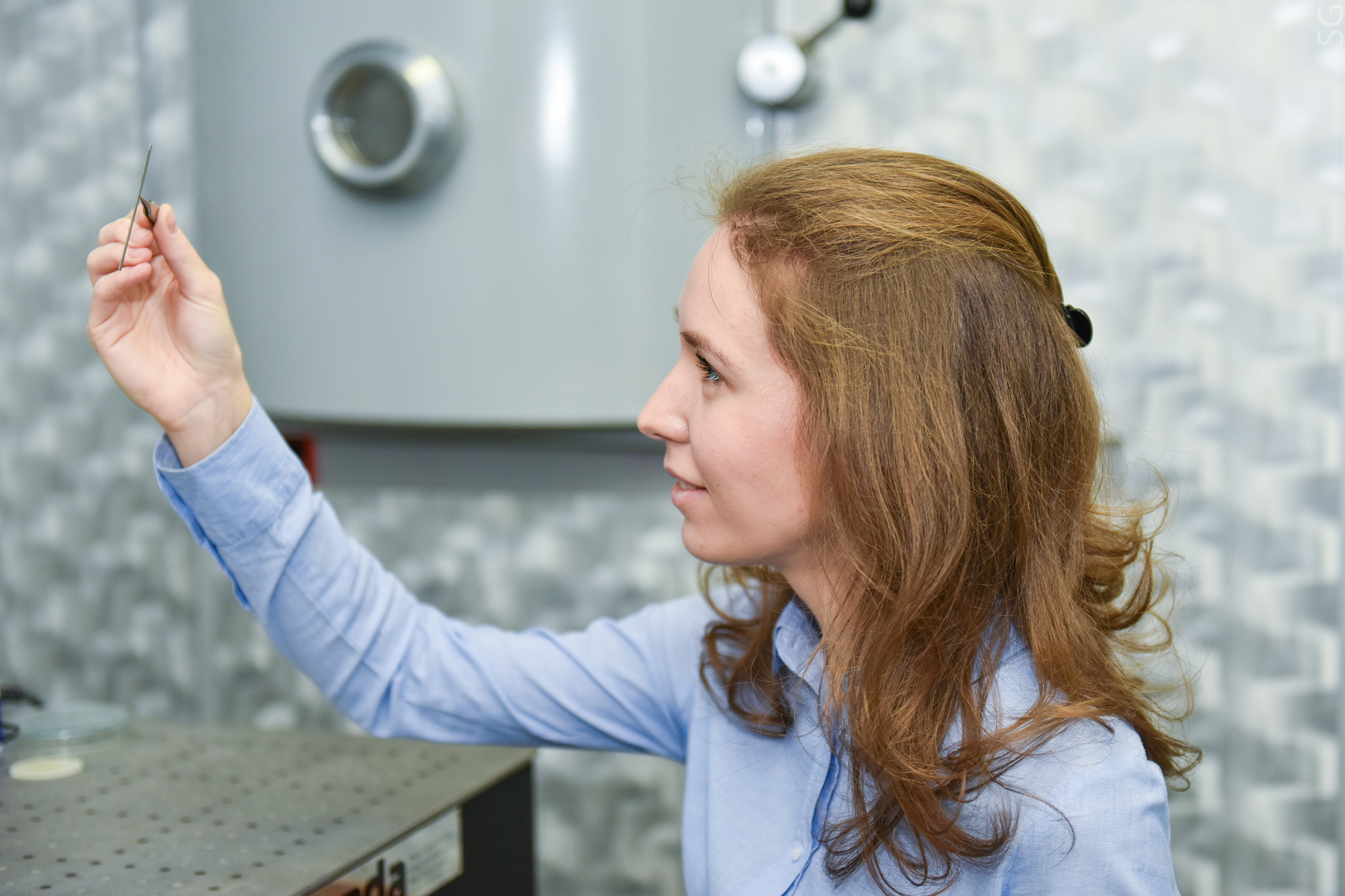
Yulia Terekhova, an employee of the Department of Materials Science of Semiconductors and Dielectrics of NITU “MISiS”, created a small scientific miracle: it improved the capabilities of the most accurate instruments in the world: scanning probe microscopes. Now they can be used to study the surfaces of substances at the atomic level with previously unattainable temperature values. What this will lead to in scientific terms, while one can only guess: since no one has seen what happens to surfaces under these conditions, the result is still unpredictable. But one thing is clear for sure: to find out what secrets the heated surface of even the most famous substances keeps in itself, all laboratories in the world will have to change the “heart” of a scanning probe microscope - a piezoelectric plate, which moves the scanning needle of the device.
Scanning probe microscopes (SPM) are research instruments that allow not only to investigate objects at the nanoscale level, but also to manipulate them with high accuracy. The principle of operation of such microscopes is based on "probing" the surface of the sample under study by a miniature needle - cantilever. It is necessary to move such a needle very precisely, over distances of the order of units of nanometers. For this purpose, special devices are used - actuators operating on the basis of the piezoelectric effect. It can be seen in piezo lighters, in which pressing a button causes a sharp deformation of the quartz crystal and the appearance of an electric spark. The reverse effect works in probe microscopes - the applied electrical voltage deforms the crystal to which the needle is attached. By changing the voltage, you can move the needle and, line by line, scan the surface in this way.
')
Now, in most scanning probe microscopes, tubes made of lead zirconate titanate (PZT) are used as a piezoelectric. He has many advantages over competitors, but he is not perfect. For example, due to such a phenomenon as a mechanical hysteresis, the cantilever can move to an unpredictable point during scanning, and the low resistance of the piezoelectric to temperature changes leads to the fact that the experimental results depend on the “weather” in the laboratory.

Julia suggested replacing the PSTN ceramics with the use of a new material developed at the materials science department of semiconductors and dielectrics - bidumenal lithium niobate single crystals to move the cantilever.
Lithium niobate itself has been known for a long time - the first samples were obtained in the 60s of the last century independently by scientists of the USSR and the USA for use in lasers and other optical devices. In addition to the outstanding optical characteristics, lithium niobate also exhibits piezoelectric properties and does not have the inherent disadvantages of PZT ceramics.
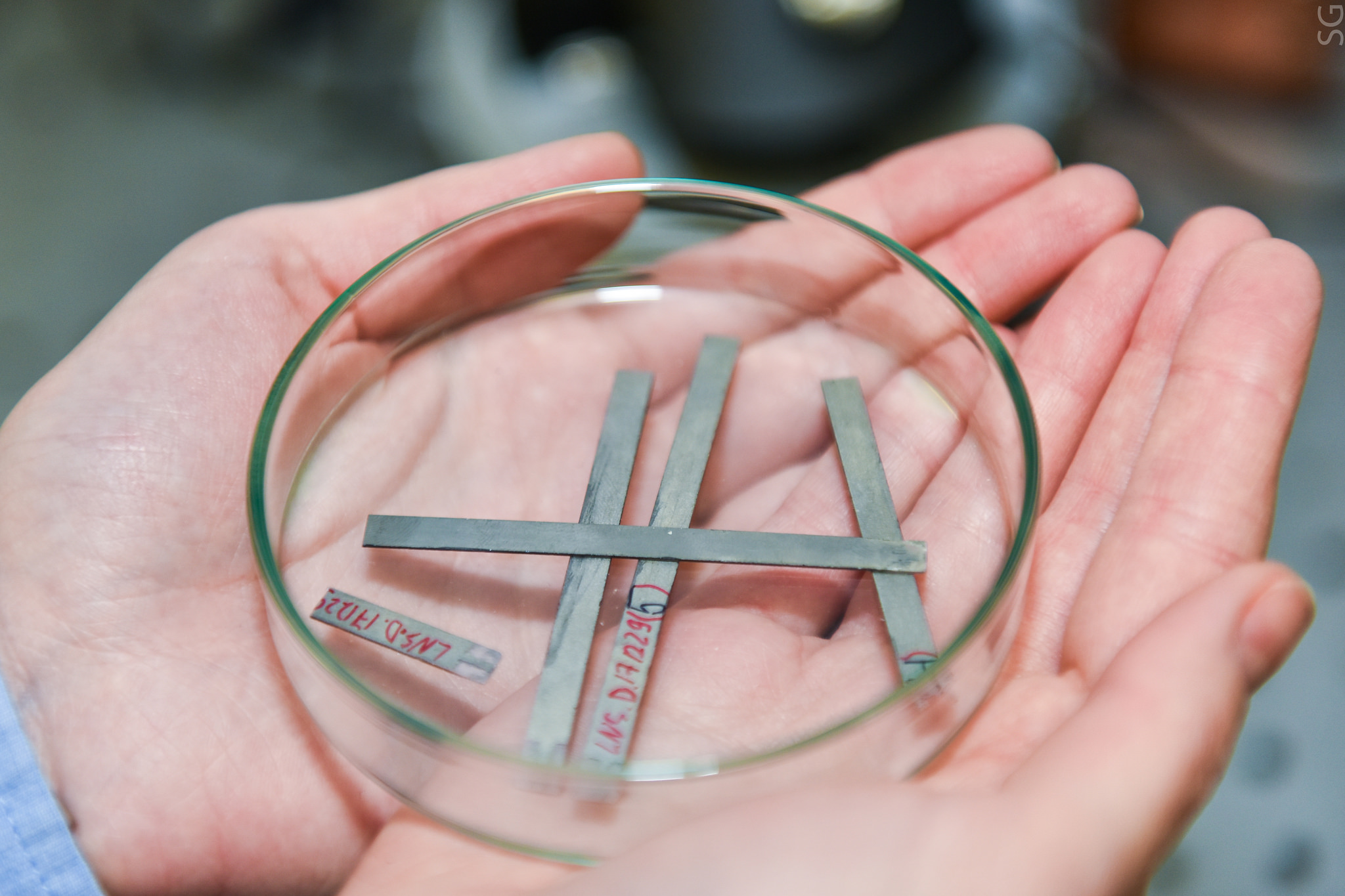
The piezoelectric characteristics of lithium niobate are an order of magnitude worse than that of piezo-ceramics, which until recently did not allow it to be used in scanning probe microscopes: too much voltage had to be applied to niobate in order to move the cantilever needle to a sufficient distance. But a group of scientists from NUST "MISiS" was able to solve this problem. A thin lithium niobate crystal plate is annealed so that it forms two regions of equal volume (domains) that are deformed differently when an electric field is applied. Such crystals are called bidomic. Having correctly selected the geometry and orientation of the plate, it was possible to obtain significant movements of the cantilever at small control voltages.
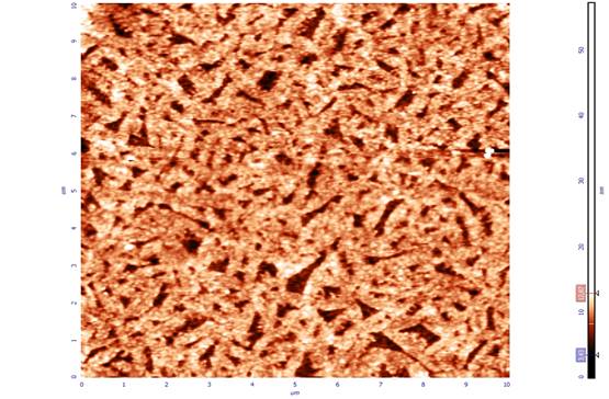
Thanks to the use of crystals from bidomene lithium niobate, the images became more clear. In addition, it became possible to investigate surfaces at temperatures unattainable for PZT ceramics. It ceases to be a piezoelectric at 150–200 ° C, and niobate retains its properties up to 450 ° C, which makes it possible to study changes in the scanned surface during heating, for example.

According to the condition of the “U.M.N.I.K.” contest of the Innovation Assistance Fund, which Yulia Terekhova won with her project, she will work on it for two years. At the moment, she is optimizing a laboratory sample of the first of its kind “core” for a microscope. The result of the study should be a ready-made device that can replace outdated displacement systems in scanning probe microscopes.
Yulia Terekhova, an employee of the Department of Materials Science of Semiconductors and Dielectrics of NITU “MISiS”, created a small scientific miracle: it improved the capabilities of the most accurate instruments in the world: scanning probe microscopes. Now they can be used to study the surfaces of substances at the atomic level with previously unattainable temperature values. What this will lead to in scientific terms, while one can only guess: since no one has seen what happens to surfaces under these conditions, the result is still unpredictable. But one thing is clear for sure: to find out what secrets the heated surface of even the most famous substances keeps in itself, all laboratories in the world will have to change the “heart” of a scanning probe microscope - a piezoelectric plate, which moves the scanning needle of the device.
Scanning probe microscopes (SPM) are research instruments that allow not only to investigate objects at the nanoscale level, but also to manipulate them with high accuracy. The principle of operation of such microscopes is based on "probing" the surface of the sample under study by a miniature needle - cantilever. It is necessary to move such a needle very precisely, over distances of the order of units of nanometers. For this purpose, special devices are used - actuators operating on the basis of the piezoelectric effect. It can be seen in piezo lighters, in which pressing a button causes a sharp deformation of the quartz crystal and the appearance of an electric spark. The reverse effect works in probe microscopes - the applied electrical voltage deforms the crystal to which the needle is attached. By changing the voltage, you can move the needle and, line by line, scan the surface in this way.
')
Now, in most scanning probe microscopes, tubes made of lead zirconate titanate (PZT) are used as a piezoelectric. He has many advantages over competitors, but he is not perfect. For example, due to such a phenomenon as a mechanical hysteresis, the cantilever can move to an unpredictable point during scanning, and the low resistance of the piezoelectric to temperature changes leads to the fact that the experimental results depend on the “weather” in the laboratory.
Julia suggested replacing the PSTN ceramics with the use of a new material developed at the materials science department of semiconductors and dielectrics - bidumenal lithium niobate single crystals to move the cantilever.
Lithium niobate itself has been known for a long time - the first samples were obtained in the 60s of the last century independently by scientists of the USSR and the USA for use in lasers and other optical devices. In addition to the outstanding optical characteristics, lithium niobate also exhibits piezoelectric properties and does not have the inherent disadvantages of PZT ceramics.
The piezoelectric characteristics of lithium niobate are an order of magnitude worse than that of piezo-ceramics, which until recently did not allow it to be used in scanning probe microscopes: too much voltage had to be applied to niobate in order to move the cantilever needle to a sufficient distance. But a group of scientists from NUST "MISiS" was able to solve this problem. A thin lithium niobate crystal plate is annealed so that it forms two regions of equal volume (domains) that are deformed differently when an electric field is applied. Such crystals are called bidomic. Having correctly selected the geometry and orientation of the plate, it was possible to obtain significant movements of the cantilever at small control voltages.
Thanks to the use of crystals from bidomene lithium niobate, the images became more clear. In addition, it became possible to investigate surfaces at temperatures unattainable for PZT ceramics. It ceases to be a piezoelectric at 150–200 ° C, and niobate retains its properties up to 450 ° C, which makes it possible to study changes in the scanned surface during heating, for example.
According to the condition of the “U.M.N.I.K.” contest of the Innovation Assistance Fund, which Yulia Terekhova won with her project, she will work on it for two years. At the moment, she is optimizing a laboratory sample of the first of its kind “core” for a microscope. The result of the study should be a ready-made device that can replace outdated displacement systems in scanning probe microscopes.
Source: https://habr.com/ru/post/410857/
All Articles