A couple more unobvious things that you might not be told before laser vision correction
Today, without the "tin", as you requested
There is already a post about how a laser cuts by creating millions of cavitation bubbles in the layer of the cornea of the eye, and analyzing telemetry from a real operation in seconds with comments from the surgeon’s actions. And the history of operations . You asked a lot of questions. Now FAQ about various related things.
- If I look away while the laser is running, what will happen?
')
You simply will not work. In fact, immediately after anesthesia, the eye is pressed against a special pneumocapture. To blink at you too will not leave because of fixing (it is not long and not for long). The only time where you can seriously disrupt the course of the operation is to jerk your head hard, with a serious willed effort pulling it out of the head restraint. In this case, the operation will stop instantly. More precisely, it will stop even before the loss of capture (details below).
- How should an operating room be prepared?
In general, it is like a normal operating room, that is, it is a room with a clean area (air filtration, excessive pressure to prevent contamination from the outside after cleaning). It is important for the procedure that microparticles of dust flying in the air do not fall between the laser lens and the eye.
- What are the chances of my head being pulled during an operation?
You have few chances, but about once every 2,000 operations the patient still manages to do this because of an external event, such as a flood or an earthquake. In serious clinics the process of operation is controlled by several people, the display is lit, the door is fixed, and everything is done to prevent this from happening. In the laser "Visumaks" there is a special function that automatically blocks the opening of the door during operation of the laser. For example, before the operation, the surgeon and the nurse alter the algorithm of actions with the patient several times. And yet, so that you are not nervous, the surgeon is constantly talking to you during the operation (surprise: most likely, when he is silent, the operation will actually end, and you will only start to think that you need to concentrate). In your hands you have a special soft toy that distracts motor control. The head lies on a special lodgement on which it is difficult to move without a conscious strong effort. In general, almost everything is thought out. Yes, you take the toy with you as a souvenir of a successful operation.
- What happens in this unlikely event if the pneumatic grip is broken?
Pneumatic gripping is designed to keep the eye soft enough, because a strong pressure on the surface changes the pressure in the eye and the geometry of the tissues. In addition, the smart bed you are lying on adjusts to micro oscillations and presses you up a little. If fixing disappears, the laser will stop the operation within a few milliseconds. This happens as follows: in the pneumatic holding there is a pressure gauge that controls, in fact, the fixation of the eye. If you pull your head, then a lot of things happen in the microcosm. First, pressure begins to fall, and then the eye loses contact with the grip. The sensor has time to turn off the laser before the eye finally loses contact and remembers the control breakpoint.
- I'm paranoid. Suppose I shook my head, the laser turned off. What's next with the eye?
Depends on where exactly the stop occurred.
- In approximately 50% of cases, it will be possible to continue normally, but the surgeon will have to make a small incision at the stopping point with a diamond scalpel (a small uncut bridge is formed there). I have 300 ReLEx operations per year, and so far there have been no cases. My partner and inventor of technology, Professor Sekundo, has been spending more — about 8 years in large quantities. With thousands of vision correction operations with such a scalpel at the ready, he has never used it during ReLEx.
- In some cases (approximately 40%), it will be necessary to make a wide cut, that is, to change the operation from SMILE to FLEX - it is more invasive, but the quality of the correction is the same. It was twice in 8 years in a German clinic, both patients did not experience complications.
- In about 10% of cases, a different type of correction will be required. We always choose the best option and stop at the slightest doubt. A micro incision in the eye itself has almost no effect, the key change in mechanics is precisely the extraction of the cut lentikula lens and the collapse of the resulting cavity.
In general, in the case of a complex stop, we recommend waiting 2 hours before the gas leaves the cavitation bubbles - this is necessary not only for the surgeon to make an informed decision and make the diagnosis again, not on the fly, but also for the patient to calm down.
- And if I shake my head not under the laser, and when will the surgeon take out the lenticle?
One such case really was during the first 500 operations in Germany: the patient got nervous and jerked, and it was at that moment that there was a sharp spatula inside the eye (the fifth generation of lasers required sharper tools — you had to cut bridges and spikes a little, now spatulas are blunt). Since then, the patient is not in danger, since the mandatory part of the technique is to grip the eye with tweezers to prevent this. Yes, by the way, in this patient, nothing terrible happened - the surgeon a little differently exfoliated the lenticle, this did not affect the quality of vision. The worst thing that can happen is that the cut will expand. It naturally increases in the radial direction - it is not very scary, just as if the surgeon made a slightly larger area to gain access to the lenticle.
- Does the laser have a seismic protection?
For 10 years of practice, we have never heard that extraneous vibrations interfere at the stage of performing a laser incision. At the stage of the surgeon's handwork, they could actually prevent, but the special emphasis under the doctor’s elbow is designed so that the patient and the surgeon’s hand become one rigidly connected system. In earthquake-prone regions there are special structures of the building in which the operating units are located.
- What is energy protection used?
The standard femtolaser VisuMax is equipped with built-in backup power batteries and has a built-in power filter. Other lasers may not have their own batteries, but have standard inputs for medical UPSs. Even if electricity is cut off throughout the clinic, the laser can complete the operation. New, however, will not start. In case of problems, there is one more level of insurance - VisuMax remembers the stopping place and is able to find it later.
- Can I come already “caught” or will I be immersed right in you in a drug sleep?
In theory, this is possible, but in practice we need a patient with responsive eyes for accurate centering (more precisely, its control). So forget about medication sleep (even light) - your 20-25 seconds of fear and discomfort are not worth the increased risk of surgery. Accordingly, for the same reason, we do not recommend using psychoactive substances before surgery. Well, do not come in altered states.
- What will I feel during the operation?
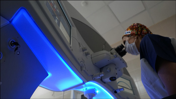
Before the operation, you will pass a triple briefing and three times listen to the story about the "green light bulb". The operation is performed on an outpatient basis, on both eyes it takes an average of 20 minutes. If you are a little nervous and nervous, this is perfectly normal. If you experience too much anxiety, you can ask for a sedative pill. Before the operation, we once again study all the data together, after which the doctor drops painkillers into the operated eye.
To prevent blinking during surgery, the eyelid is fixed with a special dilator, the second eye is closed. In order not to create the risk of external objects appearing in the laser optical zone, the patient may wash the surface of the eye and cut the protruding eyelash. Then the patient is transferred to the working position of the laser by moving the table and asked to look at the green LED that moves and becomes clearly visible. At this point, the surgeon centers the optical surface of the lens with respect to the optical axis of the eye, seeking contact between the lens and the surface of the cornea. Grabbing and fixation is performed using a special lens, adjustable in accordance with the individual characteristics of the patient's eye. Then the laser works like here on telemetry. At the end of the laser, the patient is transferred to a safe position on the table and transferred under the microscope. The doctor selects the lenticle with a spatula and removes it. In this case, the patient usually sees a bright point and feels light vibrations.
Then the patient may cry a little for a couple of hours. But not only for joy, but because of the reflex that causes tears when the eye is damaged. For some time, some people may feel a slight burning sensation in their eyes, which feels much weaker than the smoke when cooking kebabs. Most often, this stage takes place without any particular emotions. The next day, the discomfort disappears, the visual load is not limited, but it is better, in spite of this, not to strain your eyes too much with reading, telephone or movies. You can not visit the sauna for some time, so that the water does not fall into a 2.5-mm incision, which was used to access the lenticule.
- How is the laser operating mode and the shape of the cut calculated?
Firstly, one of the most important things in the preparation of an operation is accurate diagnosis and accurate measurement of the eye. Comprehensive primary examination before correction in our clinic is 16 examinations on the most modern specialized equipment under a single European protocol. This is followed by a detailed consultation with one of the professors-surgeons to discuss the correction option that is optimal for the patient, taking into account his visual needs, lifestyle and anatomy of the eye. Examination and postoperative observation of patients is carried out only by ophthalmologists with experience in the field of refractive surgery. All this is extremely important for correct calculation of laser correction parameters. Based on them, a model is set for laser automation. About this entire stage, I will tell you more in detail later. Secondly, the parameters and shape of the cut depend on the size of your myopia and astigmatism, the size and shape of the cornea, even the size of the palpebral fissure and the nose. In the case of complex cases of astigmatism, a great deal depends on the correctness of capture and alignment, but how this is done and how the methods evolved is a separate story.
Thirdly, the working mode depends on the preferences of the surgeon, on his skills and skill - we change the porosity and spot diameter to obtain the optimal quality of cuts and optimal conditions for the extraction of lenticules. Fourth, on a specific VisuMax laser machine: you need to adapt a little to your particular laser, they are slightly different in the series.
- What to do if the center of view does not coincide with the center of the pupil?
This is infrequent (about 10%), but a possible case, when the angle between the visual and anatomical axes of the eye (Kapp angle) is large. It is detected before the operation and requires a special change in the model. Here the qualification of the surgeon is extremely important. Also a topic for a separate story.
- How about the corrections after SMILE?
In our practice (German and Russian), not a single patient had the need for an additional correction. But technologically there are special methods that allow for additional correction. How exactly - decide the doctor. At the European Congress in Copenhagen this year, Dr. Dan Renstein gave a report in which he listed 5 different methods of additional correction in different cases. So, if the patient suddenly needs, there is plenty to choose from. After SMILE, the quality of optics on 6th generation lasers is consistently higher than after LASIK or femtoLASIK, the probability of side optical effects is significantly less. When we are planning a laser correction, we expect that this procedure is done only once in a lifetime. The percentage of the correction on the generalized international statistics for SMILE is the lowest today - about 0.5%. In specialized clinics with experienced doctors, it is significantly less. I repeat, we didn’t have them specifically, but the likelihood cannot be ruled out, so this is rather a question of experience and luck.
- Why do I need to get lenticular, and not evaporate it inside the eye?
In theory, of course, you can try to evaporate the layer after layer of lenticular inside the cornea, without “opening” it at all. The difficulty is that it will require the removal of decay products from the eye, and the exchange of fluids there is very, very slow. According to preliminary calculations, in theory, it is possible to make 0.25 diopters per week, plus a perfect positioning system will not yet be needed to continue the work started last time. Well, either technical tags right in the eye, which is also not the best idea. In practice, the restriction is even simpler: no patient will pay 10–20 times per operation (and the price will be slightly lower than with the usual ReLEx) and go to it for half a year every week. Therefore, while this is an idea for the future.
- What will happen when nanolasers appear?
Now nanosecond lasers are undergoing clinical trials. Their use will allow working at higher frequencies with a slightly lower heat transfer into the fabric, but before the first practical operations “into combat” there is still about 5–7 years at least, rather, all 10. This is a quantitative growth of several percent, rather than qualitative, in the technology of operations in the eyes now, little will change fundamentally.
- Why is it now often possible to meet proposals for laser vision correction at very low prices, what is there to save?
Now there are still a lot of clinics that work on morally and physically outdated equipment for diagnosis and surgery. And sometimes without the necessary diagnostic equipment. There are even such clinics that call themselves "premium", and the equipment of the past and before last generations. Hence the complaints of patients in the postoperative period on the quality of vision and other problems. Older laser systems are not capable of solving problems for which modern lasers are designed. Therefore, when choosing a clinic, ask what laser device will be used and what year it is manufactured.
- How much stronger is the cornea weakened during the LASIK procedure than with ReLEx?
So far, the understanding is intuitive: ReLEx is better. A practical report on a multi-year study on biomechanics will be published in 7 months. Then we give the numbers and facts. In general, you can no less intuitively compare the results on the loss of innervation and other side effects such as keratoconus - in the case of LASIK / FLEX-methods there are many times more. But, I repeat, it is better to discuss this in numbers after the publication of 10-year studies, which are simply not there yet. Quite briefly - the FDA has the most stringent requirements and always insists on conducting its own independent research, but it found SMILE suitable for use in US clinics as one of the main methods. In Russian Wikipedia there are statistics on the side effects of various operations with confirming links .
- Why is ReLEx made only by femtosecond lasers?
At Lasik, two lasers are used: femtosecond to remove the “cap”, and then excimer to evaporate a wide lens. For precise minimally invasive (and transmitting little heat to the bowman membrane and below) operations, lasers with a frequency of about 500 KHz and very precise focusing are needed — traditional excimer ones are not suitable. Currently, only Zeiss optics provide the necessary accuracy, that is, lasers of the VisuMax series (the old models are most likely familiar to you). 13 microns thickness is one diopter. This accuracy was achieved by combining the actual laser tube from the industry (initially, seemingly cosmic) with Zeiss lenses. It was the latter who created focusing optics for medicine.
- What you need to know about the cornea and what features of the regeneration of each layer?
The cornea is the part of the eye that you can easily see: a convex transparent part that contacts the air. The usual diameter is 10–12 mm for people of any race. In the center, the thickness of this convex-concave lens is 520–560 microns, with an edge of about 1 millimeter (all sizes are average, there are thin and very thin corneas). The cornea has 5 layers. Here is a schematic diagram:

But on a scale from Commons:

1. corneal epithelium; 2. Bowman's membrane; 3. corneal stroma; 4. Descemetov shell; 5. corneal endothelium.
The corneal epithelium is a multi-layered flat tissue and accounts for about 10% of the entire thickness of the cornea. Corneal epithelial cells are located in 5–7 rows. The corneal epithelium performs a mechanical protective function, since it prevents microorganisms and foreign bodies from penetrating inside the eye; biological protective function, because it contains cells that are involved in the immune response, the optical function - the mucin of the tear film fills all uneven elements in the surface layer, which provides a smooth, transparent surface for the passage and refraction of light rays; membrane function - the corneal epithelium is a biological membrane through which certain substances can penetrate. Like the normal epithelium of the skin, it regenerates well, and during PRK surgery it is removed to gain access deeper. Within a few days, it is restored.
The second layer is the Bowman's membrane. This is an incredibly thin and important layer immediately under the epithelium. Bowman's membrane is located under the basement membrane, has a thickness of 12 microns and does not contain cells. The Bowman's membrane consists of randomly arranged collagen fibrils. It has a smooth front surface and a back surface for smoothing the non-uniform relief of the stroma, which ensures the transparency of the cornea.
The Bowman's membrane cannot be restored after damage, therefore, after corneal damage in this part, scars are formed at the site of defects and the transparency of the cornea in these areas is disturbed - turbidity is formed. When laser correction lenses are formed deeper. However, at any cut through the bowman's membrane, we cut the nerve endings. In a PRK operation, it is removed to gain access to the stroma.
Accordingly, with minimally invasive removal of lenticules with ReLEx, the cut there is only one short by 2.5 mm, and not nearly the entire circumference, as in the case of LASIK / FLEX-like methods. It is the injuries of the Bowman's membrane that violate the epithelialization of the eye, innervation and give other side effects.
The next part of the cornea is the stroma. This is where the main work goes. Fabric - collagen filaments impregnated with hyaluronic acid. When magnified, they resemble ropes, such as in section:

The stroma is the main part of the cornea and occupies approximately 90% of its thickness. Corneal stroma consists of parallel plates. Plates are formed from collagen fibrils. Collagen provides transparency of the cornea and its strength. In the stroma of the cornea there are two main parts: the anterior stroma of the cornea and the posterior stroma of the cornea. The anterior stroma is looser and consists of thinner plates; the posterior stroma has a denser and more compact structure.
Stroma regeneration is carried out by keratocyte cells, which are capable of synthesizing collagen and, due to this, maintain an optimal level of collagen fibers and extracellular matrix.
These same ropes can perfectly coalesce, if they are stuck in one another (with the formation of nodes-adhesions, which interferes with visual acuity), but at the same time, being put on each other overlap (that is, from different angles), do not form these nodes, but simply interlock. During the laser correction ReLEx SMILE we cut the lens in this layer and pull it out. After the operation, the cavity in the cornea closes - the “ropes” lie on top of each other, but at the sites of the incisions a clear boundary does not form from the joints, that is, everything remains transparent (splices of single collegan filaments occur at the border of the lens, that is, in outer diameter). The frame is supported as usual - the bowman membrane stretched from above and the lower layers.
The following two layers - Descemet's membrane and endothelium - are almost not interested in laser correction, since we are not affected by these operations. This is actually a soft and rigid boundary of the body, a kind of standard “casing” for the body.
- What will happen after the operation?
On the day of surgery, most often after a couple of hours, you hardly feel any discomfort and it disappears completely the next morning. The main limitation after surgery is not to rub the eyes with your hands. During the week after the surgery, you need to bury the eye drops obtained in the clinic. If necessary, we will also provide preparations of artificial tears.
Visual acuity in the first days may vary somewhat. In order for the surfaces to come together after extracting the lenticules, a slightly longer process of visual rehabilitation is needed than with methods that imply “full opening” of the cornea of the eye. It takes time for the eye to learn to focus on close objects in order to make the right muscles work. We conduct examinations to check the results of the correction every other day, a few days later and a few more weeks after the operation.
After two or three days, you can get back to work and play sports. In general, the next day after surgery, you can safely lead a normal life (ride a bike, stoop, etc.) - there is no risk of flap-flap displacement, as with other techniques. The visual load from the day after the operation is not limited, but it is better not to abuse the laptop, books and tablet during the first week. However, swimming and visiting the sauna should be abandoned for a couple of weeks in order to reduce the risk of infection through an incision in the cornea.
If we compare it with LASIK, then the recovery of vision is more gradual, within a week, but there are much less restrictions on physical exertion. , , , — . , , , , — , . SMILE . LASIK/FLEX- , — .
Source: https://habr.com/ru/post/400183/
All Articles