DNA is only half the chromosome volume. Everything else is a shell of unknown functionality.
Three-dimensional model of the 4th human chromosome
At school, we were taught that the nucleus of each cell contains filamentary structures called chromosomes, in which genes are stored — units of heredity, arranged in a linear fashion. Genes are encoded as part of the DNA macromolecule. But everything is not as simple as it seems.
From the very moment of its discovery in 1882, the chromosomes underwent a thorough and close study, including with the help of optical and electron microscopes. Surprisingly, scientists are still not able to clearly understand how their structure is organized.
For decades, scientists have concentrated their efforts on the study of mainly chromatin. This is the main functional substance of chromosomes, which is a complex of DNA, RNA and proteins. It is within the chromatin that the realization of genetic information occurs, as well as the replication and repair of DNA.
')
One of the main mysteries is how the packing (folding) of chromatin occurs. For a long time it was assumed that the packing happens randomly, but lately other theories have appeared. Some scientists have suggested that packaging occurs following a polymer melt pattern . There are opinions that chromosomes pass through a chain of interrelated packaging processes, from helical winding around the nucleosome , to a solenoidal 30 nm fiber, and then to a larger helix . In the end, there is a third class of theories that suggest that chromosomes consist of chromatin loops held back by non-histone proteins .
The last of the listed models of the chromosome structure has recently received additional confirmation. In 2013, advanced microscopy techniques clearly showed how a linear matrix of chromatin loops is formed in the cell nucleus (see the work of Natalia Naumova of the University of Massachusetts and colleagues, published in the journal Science ). The video shows more how self-organization of chromosomes occurs.
Organization of mitotic chromosomes (accompanying material to the article by Natalia Naumova and her colleagues in 2013)
However, all these chromatin studies by and large ignored the thin surface layer, which was discovered on chromosomes by the method of classical microscopy in 1968 . This peripheral layer was investigated poorly, and its composition and structure remained virtually unknown. By default, it was assumed that it was just some kind of amorphous mass that stuck to the chromosomes.
The problem is that we can study the chromosomes only in certain conditions, so we do not have a clear idea of how they actually look.
A group of British scientists from the University of Edinburgh has been studying the outer covering of chromosomes for several years. Several years ago, they proved that assembling the outer part of the chromosome necessarily requires the presence of Ki-67 protein. Scientists have suggested that chromosome plating is made with the participation of this protein.
A new study conducted by a group of scientists, contains the results of 3D-modeling of the structure of the chromosome, as well as a description of the shell. According to scientists, this material amounts to 47% of the chromosome volume. At the same time, the functionality of this material is still unclear.
Presumably, individual chromosomes are isolated from each other due to the membrane during the key process of cell division. Probably, this material also helps to avoid mistakes when dividing cells. It is known that certain types of cancerous tumors and congenital human diseases are associated with such an erroneous division.
For the first time in the history of science, scientists have compiled detailed 3D models of all 46 human chromosomes. This was achieved through the use of the new 3-D-CLEM method, which combines optical and electron microscopy and allowed us to remove chromosomes with unprecedented resolution.

Schematic description of all stages of the 3-D-CLEM process, which takes from 5.5 to 19.5 days
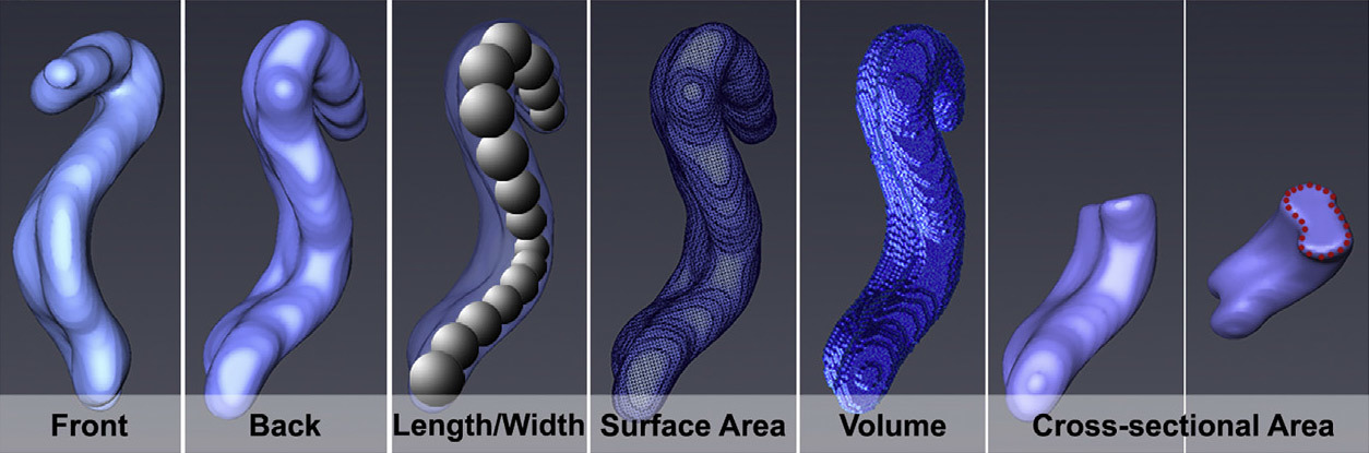
As a result, scientists have carried out a simulation of the length, width, surface area, volume and density of DNA packaging in chromosomes.

“The visualization method we developed for studying chromosomes is truly revolutionary,” says Dr. Daniel Booth of the University of Edinburgh. - The first studied structure of all 46 human chromosomes led us to rethink the idea that they are almost entirely composed of chromatin. This assumption remained unchanged for almost 100 years. ”
In fact, chromatin is only from 53% to 70% in the composition of chromosomes. All the rest is a shell that turned out to be much thicker than imagined.
Understanding the structure of chromosomes and the shell, as well as the process of division will help to study and prevent the development of certain diseases.
The scientific work was published on November 10, 2016 in the journal Molecular Cell (doi: 10.1016 / j.molcel.2016.10.009, pdf ).
Source: https://habr.com/ru/post/399449/
All Articles