For the first time, a full atlas of the human brain with a cellular resolution of 1 µm / pixel
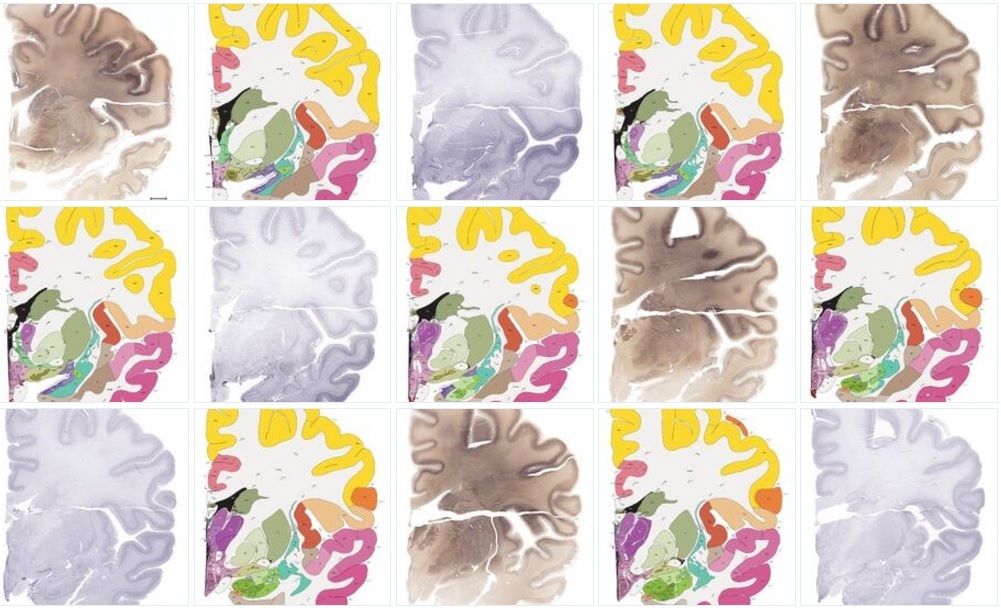
Multiple images from the atlas of the human brain. Image: Allen Institute for Brain Science
Knowledge of the detailed anatomical structure of the human brain is crucial for understanding its functionality. Existing reference atlases are not of high quality: they have relatively low resolution or they are incomplete, or the structure annotations are not enough. For a long time, the atlases of the human brain were inferior to the atlases of the brain of worms, flies and mice in quality, spatial resolution and completeness. This is due to technical limitations due to the enormous size and complexity of the human brain. What can I say, if in medicine still often used atlases a hundred years ago.
It’s good that there are patrons in the world, such as Microsoft co-founder Paul Allen. Half a billion dollars invested in a research project to study the human brain, brought the result.
Scientists from the Allen Institute for Neuroscience (Allen Institute for Brain Science) have filled a large scientific gap - and have prepared the most complete and detailed digital atlas of the human brain today. It was compiled by neuroimaging, a high-resolution histological study and a chemoarchitecture of the brain of a 34-year-old woman (as is known, the woman’s brain is not structurally and functionally different from the man’s).
')
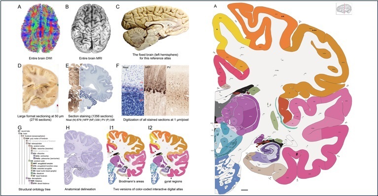
Magnetic resonance imaging, diffusion-weighted imaging (diffusion MRI) and 1356 large anatomical plates the size of the brain hemisphere for immunohistochemistry (IHH) and Nissl’s cellular resolution, that is, 1 μm / pixel, were used to compile the atlas.
Earlier this year, the creation of an atlas of the human brain was announced by researchers at the Human Connectome Project . They compiled MRI images from 210 adults. But such a picture cannot even compare with the detailed atlas of a single woman at the cellular level, which was compiled at the Allen Institute on neuroscience.
“This is the most structurally complete atlas of today, and we hope that it will serve as a reference guide for the human brain in various scientific disciplines,” says Ed Lein, Ph.D. and Allen’s researcher in neuroscience. In the future, this atlas will be useful as a basic reference for overlaying other maps of the human brain, including functional maps or maps of the cellular composition of brain regions. This is a kind of unified framework for integrating a multitude of scientific studies, combining different types of data on the human brain.
The atlas includes fully annotated images of 862 structures of the brain, including 117 paths (neural paths) in the white matter and several new cyto- and chemo-architecturally selected structures. These annotations, obtained from anatomical plates and Nissl studies, were also transferred to the corresponding MRI images, where the brain was still not completely cut.
Atlas of the human brain. Illustration: Allen Institute for Brain Science
In the new cortex (neocortex), researchers have demarcated separate areas: grooves, convolutions, and modified Brodmann fields in order to link macroscopic anatomical parcelations and microscopic chemoarchitectural parcelations into a single structure.
In order to create a complete ontology of the brain and precisely distinguish individual regions in cross sections, scientists at the Allen Institute had to develop a new scanner that could scan thin sections of tissue the size of the full hemisphere of the brain with micrometer resolution.
Scientists have done a great job. The atlas of the woman’s brain created by them is so detailed that it makes it possible to quite accurately determine the corresponding structural areas in the brain of other people according to MRI data.
The interactive digital brain atlas, compiled by the Allen Institute for Neuroscience, is freely available to the scientific community around the world. On the site, it is integrated with existing gene expression atlases compiled at the same institute.
Digital atlas in its format is very different from the classic atlases. The interactive format allows you to "travel" in the brain, change the scale from macroscopic to microscopic - from whole parts of the brain to specific neurons. In this case, the atlas contains absolutely accurate information from scientific papers that have been reviewed.
The Allen Neuroscience Institute is a private research institution that was founded by Paul Allen, one of the creators of Microsoft. The philanthropist has invested more than half a billion dollars in a scientific project to compile an atlas of the human brain. This project is called the medical "Project Manhattan" .
The medical “Project Manhattan” should help to understand how a person’s thinking process occurs: “I, as a former programmer, are very interested in how the human brain works, how information processing actually takes place,” said Allen in an interview. - Even quite a bit deep into neurology, you begin to understand that everything is interconnected. That is, the brain is trying to use everything that is possible: sight, hearing, experience of the past. The brain uses all this to calculate what the animal (be it a mouse or a man) should do next. ”
Ideally, the atlas of the human brain will help to understand the most important thing - what is consciousness.
The scientific article was published in the Journal of Comparative Neurology (doi: 10.1002 / cne.24080). Actually, the 350-page atlas occupied the entire issue of this magazine.
Source: https://habr.com/ru/post/397783/
All Articles