What can be diagnosed in the eye for a hundred rubles
Here, Milfgard recently did a post about glasses, and somehow the question “But why pay for diagnostics in the clinic, when in any optics they make it for you for a hundred rubles.” Or free of charge altogether. ”After a five-minute stupor and a couple of nervously drunk cups of coffee, it came to the realization that many simply cannot imagine what the complete eye examination consists of. In short, I’ll tell you how everything should look if there are good doctors and the right equipment in the clinic. There are a lot of photos under the cut, we tried to show what the patient usually doesn’t see - directly the working interfaces of the devices with which the doctor works. And at the end of the publication we prepared a small nice bonus.
You need to talk with the patient
Oddly enough, but this is a key stage of the study. Banal finding out the place of work and habits can give a lot of information for the doctor. For example, from the same programmers and admins, he immediately expects problems with accommodation due to chronic overwork. And with a high degree of probability xerophthalmia will arrive - dry cornea and conjunctiva of the eye. At the same time, they are more likely to sincerely believe that their myopia is progressing or that it should be so. Symptoms are the same, but accommodative disturbances can be corrected with proper therapy. The choice of treatment tactics also largely depends on this stage - drops can be recommended to one patient, glasses with progressive lenses to another, and a third one, for example, replacing the lens with a multifocal lens.
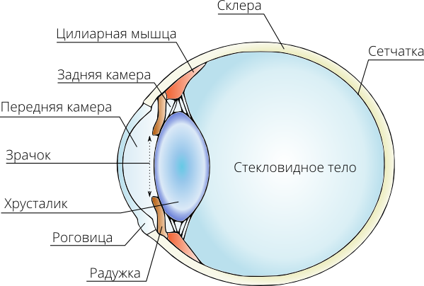
It is here that the first logical chains begin to line up. For example, the patient slouches, is clearly short-sighted, and wears a bandage on his wrist due to a ligament injury. In this case, the dislocation for him is a familiar thing. Everything, we can safely assume a genetic defect with connective tissue dysplasia. Collagen, for example, is synthesized crookedly and does not perform its supporting functions. The sclera of the eye was too elastic and the eye stretched along the optical axis during the growth period. Subsequently, he can continue to crawl a little. At the same time, after the examination, we can send him to look for
')
We expand the pupil
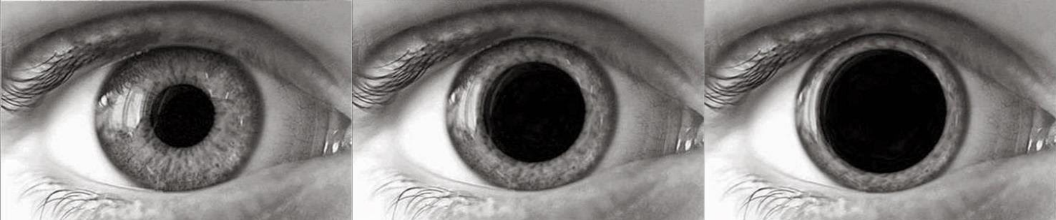
Mandatory phase with extremely rare exceptions due to contraindications. In order to expand the pupils are drugs holinoblokatory. They block the conduction of a nerve impulse in cholinergic synapses. As a result, they cause dilated pupils and temporary paralysis of the ciliary muscle, which is responsible for accommodation (changing the shape of the lens during focusing).

Meet - belladonna, she is a belle and mad cherry. The leaves contain a large amount of atropine. Very popular was a plant among girls in Italy. They buried the belladonna juice in the eyes, the pupils dilated, visually enlarging the eyes. In the daily work of the clinic atropine is used quite rarely because of its severity. As a rule, it reliably turns off the ciliary muscle and dilates the pupil by about a week. And this is an obvious search, if we just want to make a qualitative diagnosis. Therefore, usually used drugs from the same group, but with a short period of action.
These drugs, we achieve several effects. Forced dilation of the pupil, which is very similar to the diaphragm of the camera, allows in detail to consider the “sensor” - the retina of the eye and the fundus of the eye. It is also very important for a full inspection of the lens. The second important effect is paresis of the ciliary muscle and accommodation switching off. For example, a person comes to the clinic with complaints that he does not see well in the distance. We put him behind the apparatus for determining the refraction - avtorefkeratometr. He thinks and gives us the result in -4.5 D. Drip the drug, wait a few minutes and repeat again. And the result is already -2.0 D. This is true myopia. The remaining 2.5 diopters are a consequence of accommodation disturbance that can be cured. By the way, often the same holinoblockers are a part of therapy.
Autorefkeratometer

The same, all familiar thing, which shows a balloon and a house in the process of research. The main task is to establish the features of refraction of a particular patient. Returning to the title of the post, this is the very equipment that is in all optics and most often the only thing that is included in the same free examination. I planted a man, took aim, pressed a button, got a result and calmed down on it. Therefore, all these examinations for 100 rubles are so superficial. However, this does not prevent the device from being absolutely indispensable in a comprehensive study of the patient's vision.
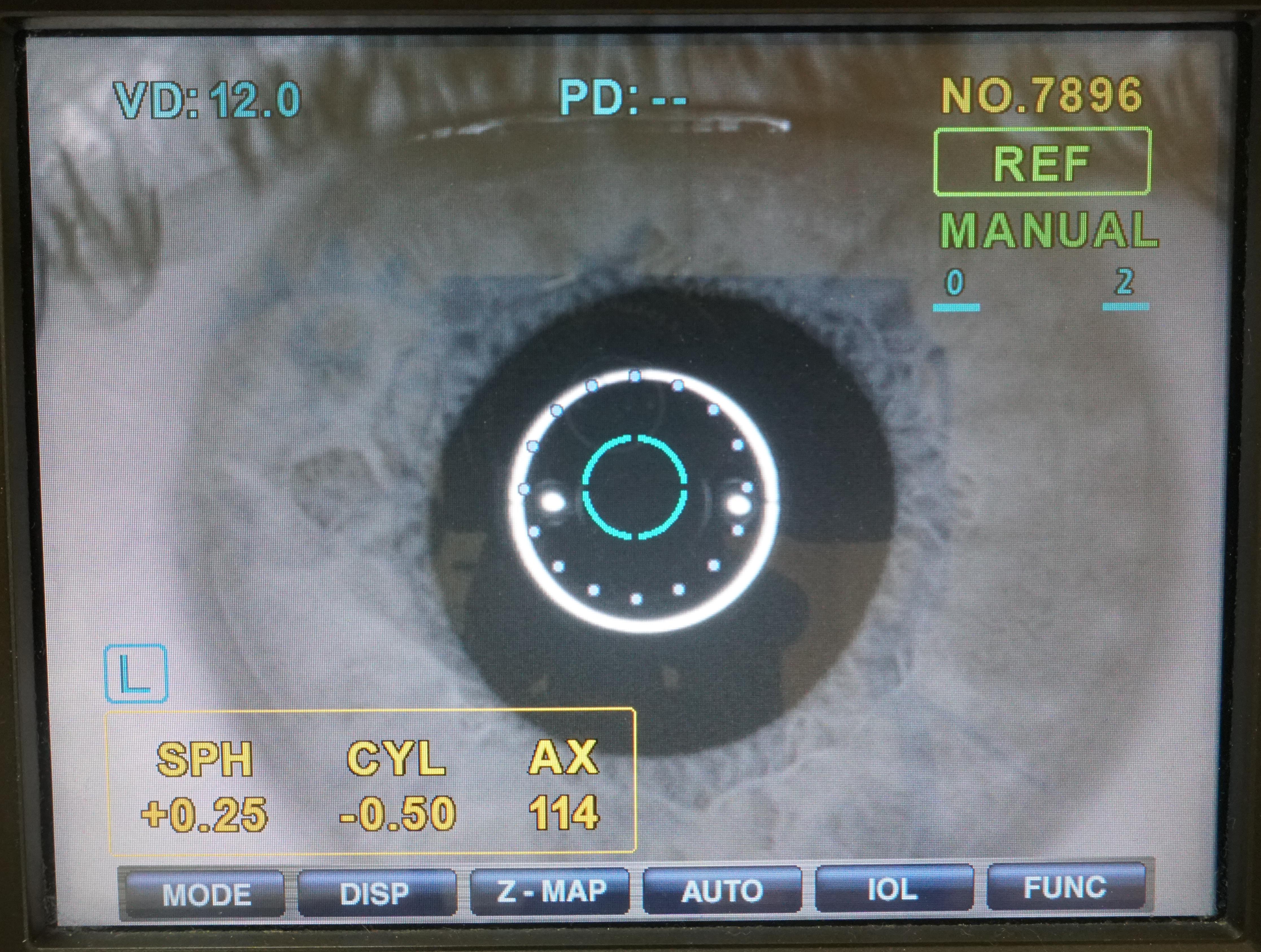
Interface autorefkeratometra. A thousand regrets that were filmed practically on sneakers, but you cannot do native unloading from the device
The device emits a beam of infrared radiation that passes through the pupil and is reflected from the retina. Special sensors carry out the registration of the image before and after the reflection moment, that is, at the entrance to the eye and at the exit from it. Further, the computer analyzes this data and provides the values on paper. The patient himself during the study sees the image, usually a balloon or a Christmas tree, which seems to be in the distance. Then, this image begins to come closer and becomes clear. At this time, the refractometer determines where the focal point of the eye is, comparing the point of a clear image, visible to the patient, with a built-in standard. Thus, we determine the refraction and detect the presence of myopia, hyperopia, astigmatism.
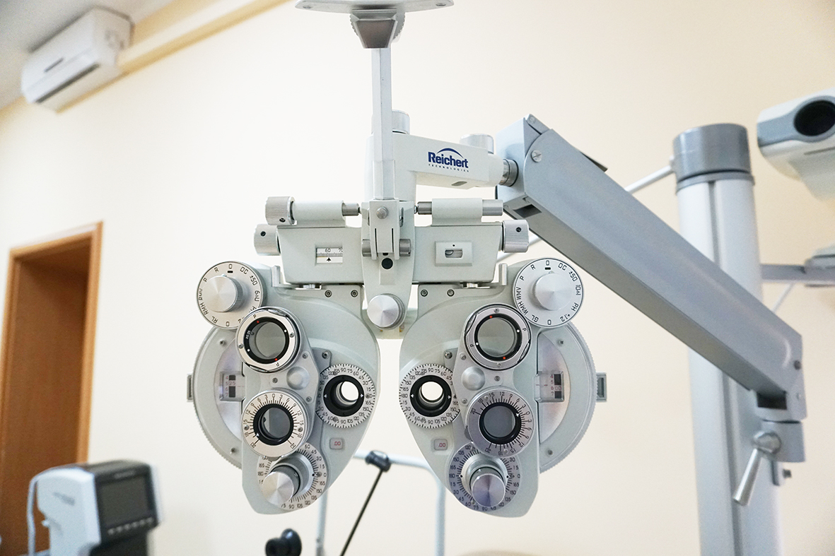
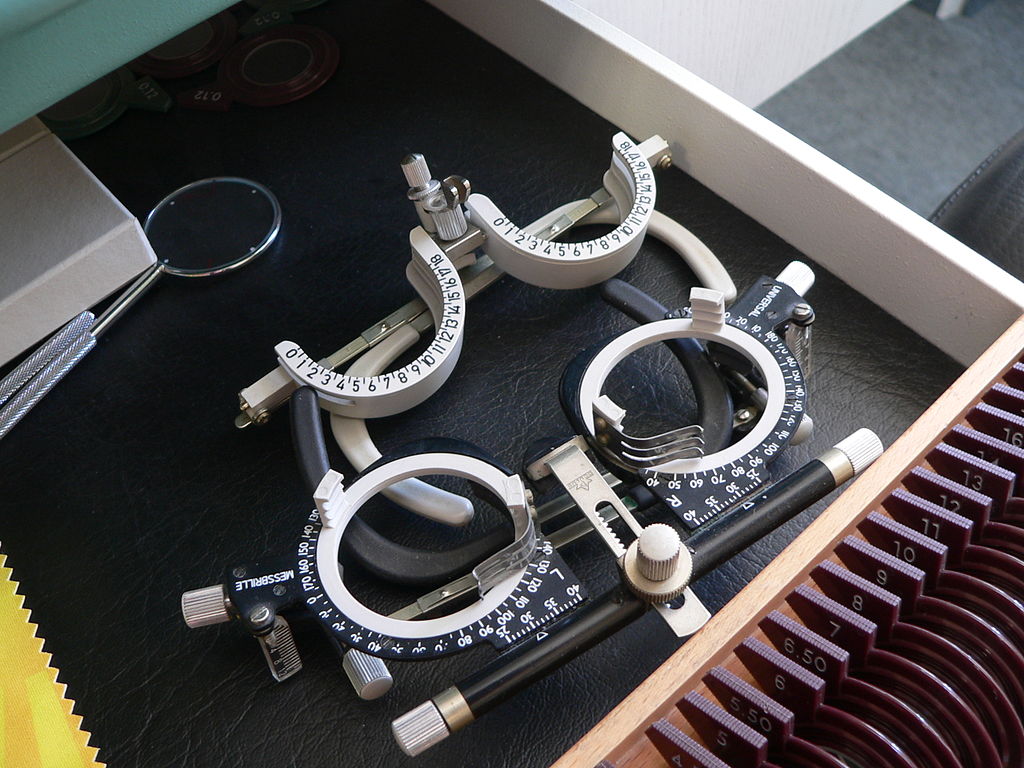
Voropter

There is something steampunk in it
This device is used to determine visual acuity and to select the right optics for glasses. The point is that the readings of the autorefkeratometer are good as a reference point when selecting lenses, but it is highly undesirable to make glasses at once based on its results. In fact, the patient’s subjective tolerance of the selected prescription plays a key role. This is where the equipment helps.

Stationary phoropters are good in their accuracy, lack of backlash, which can spoil everything during subtle manipulations, such as defining axes during astigmatism. With phoropter lens sets are used in increments of 0.25 diopters. Plus, you also need a calibrated projector of signs that forms an image of a wall or screen with given angular dimensions.
Another thing that I really want to mention. Traditional rake such. It is very important not to pick up glasses immediately after the "express diagnosis" in optics. In this case, you risk making them too strong due to the lack of separation of the component of true myopia from temporary accommodation problems. Everything will be seen sharply and clearly, but after a while the lens muscle will send you very far. There will be severe consequences of chronic overwork and headaches. Recover later will not be easy.
And sometimes you can find such small options. a source
Pneumatic pressure monitor


Very important device. It allows you to quickly and painlessly determine intraocular pressure. This indicator is important to us, because we are always afraid to miss the initial stage of glaucoma. The disease progresses gradually and due to chronically increased pressure causes dieback (atrophy) of the optic nerve. The saddest thing is that people often do not notice problems for a long time until the world around them narrows to the tank gap. A slight increase in pressure for the eye imperceptibly, and optic nerve atrophy sneaks up unnoticed, from the periphery of the visual field. Vision remains high for a long time. And we do not notice the problem on time because of our overly intelligent brain, in particular the occipital lobe, which is responsible for vision.
Our brain has a whole bunch of advanced algorithms for processing raw images from the visual apparatus. There is the “drawing” of the left frame picture from the right pieces, and refinement based on the previous frames, when the look slid over different parts of the object. Such a kind of video codec with some redundancy. From an evolutionary point of view, everything is fine here. A conditional primate is much more likely to survive if it can distinguish a banana from a leopard in time, even with an affected retina. There is no one to treat it anyway, no one will even attach the plantain. And for humans, these algorithms play a cruel joke, masking the problem until the picture crumbles away at all.
And at the late stage of glaucoma, at best, we can stop the process, but not return the lost. Therefore, we examine everyone, even those who have not made any complaints about this.
The principle of operation of the device is quite simple. Imagine that you have a ball and you put a kilogram of weights on it. If the pressure in the ball is large, then it is almost not deformed by the weight of the load. With low pressure, it significantly flattened. Pneumothometer uses the same principle. He pokes a dosed stream of air into the eye and photographs the result in the infrared spectrum. The more the cornea is pressed in, the lower the pressure. The method has a certain error, but on the whole is quite humane. No injuries and discomfort. Maklakov tonometry looks a little more unpleasant. There, in general, a small weight of paint is placed directly on the eye and the diameter of the print is measured. And abroad, Goldman’s applanation tonometer with a similar principle is more often used. Although this manipulation is generally fairly well tolerated. The contact method is more accurate, but really necessary for a very small percentage of people. The rest is fast and comfortable screening at the pneumotonometer. By the way, I know the real story when a man, after such a tonometry, made an imprint of his eye on the official press of his company.
Slit lamp


Using a slit lamp, we can see the optical cut of the cornea, look into the depths of the eye. The narrow light beam of the lamp, in contrast to the frontal lighting, gives us more detailed information about the state of the eye structures. To record the results of the observation lamp is equipped with a camera. Consider the internal structure of the eye helps high-diopter lens. With it, we can see the retina, optic nerve and fundus vessels. Naturally, the pupil for this must be expanded.
Optical coherence tomography

Another photo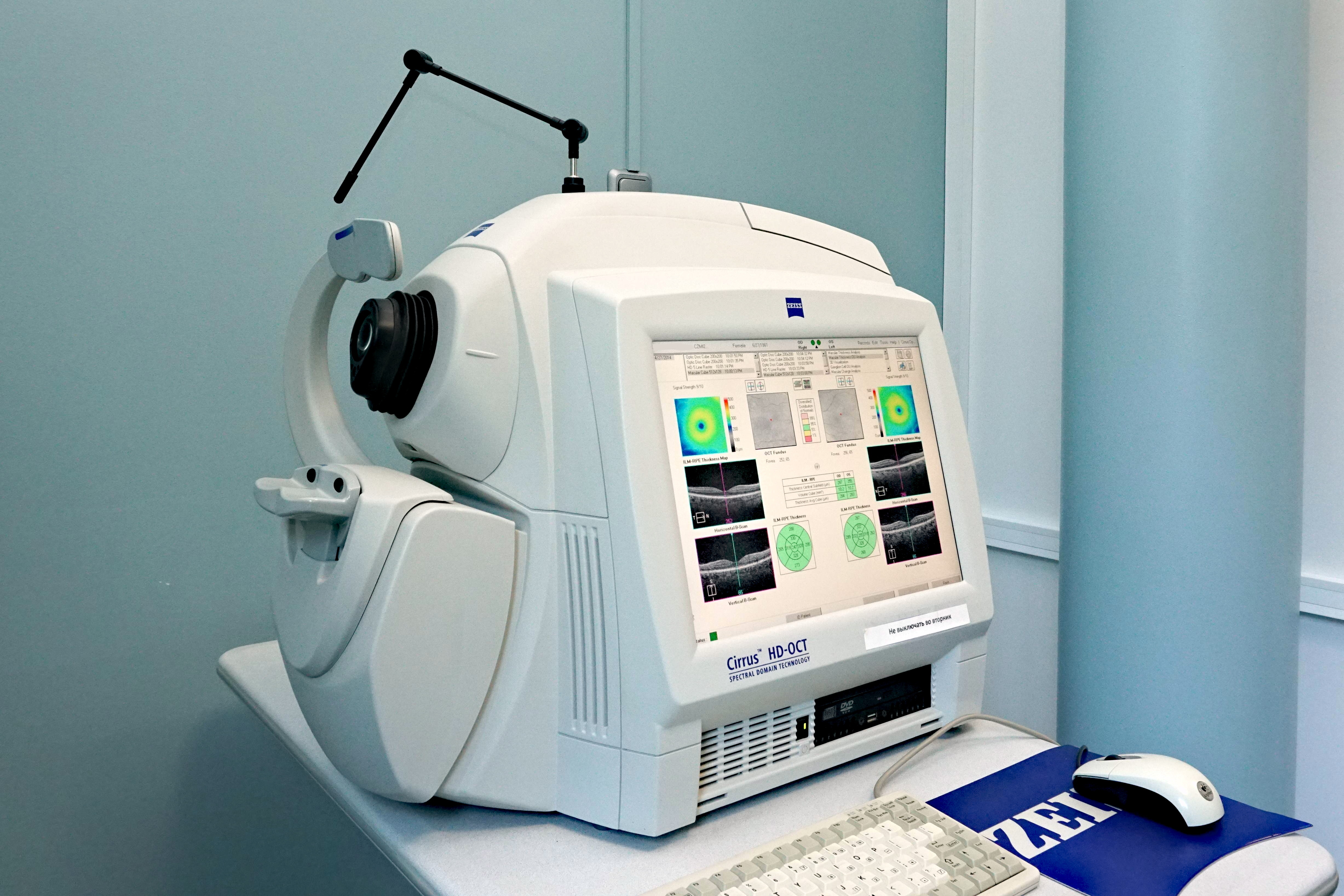
The coolest thing that allows us to get a detailed cross-section of the posterior segment of the eye in a non-contact way. We are always very afraid of possible complications with the retina, arising as a result of myopia. The whole nasty thing of myopia is that the sclera can stretch almost unlimitedly. Yes, it's sad when the eye is severely deformed and it turned out -12D at the exit. But this is less than half the problem. The sclera is primarily a connective tissue, it is elastic. But the retina and the vascular layer, which are attached to it from the inside - no. As a result, we get a stretched spring, which is ready to “shoot” with detachment or tearing of the retina in the event of any unsuccessful sneeze.
Predicting such problems at an early stage allows you to take certain preventive measures and prevent serious consequences for the patient's vision. Therefore, we treat patients with myopia especially carefully.
The resolution for such a scan is about 3 micrometers. The principle of operation of the tomograph is based on the fact that different structures of the eye reflect light differently. Using the Michelson interferometer, the wavefront velocity through the eye tissue and its intensity are measured.
The retina is illuminated with an infrared beam with a wavelength of 830 nm, and for the diagnosis of the anterior segment of the eye is used 1310 nm. This is due to their different depth of penetration. The beam is divided into two beams, one of which is reflected from a special mirror, and the second from the tissues under study. From their interference pattern, a synthetic image is obtained. The software draws a picture in conventional colors, where the ability of the fabric to reflect light increases in the red direction. A normal vitreous body should look optically transparent, that is, almost black.

Retinal tearing in the macula area
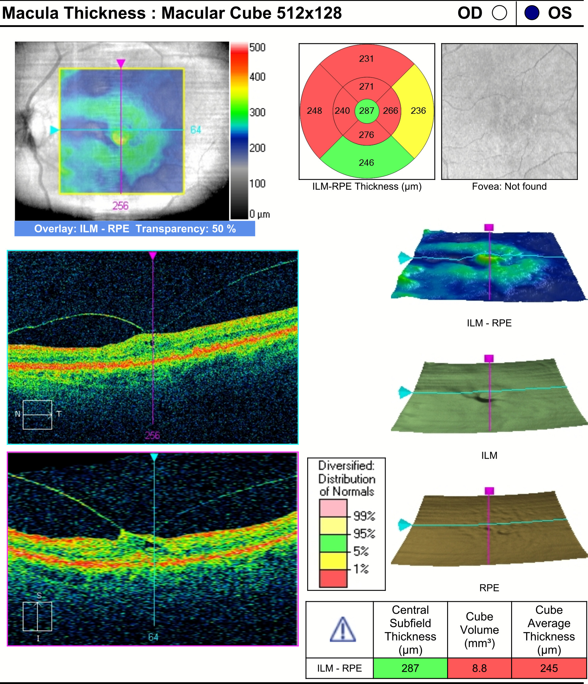
Vitreous body attached to the retina and pulls it to itself (vitreo-macular traction syndrome)
Diabetic Macular Edema
Keratotopograph
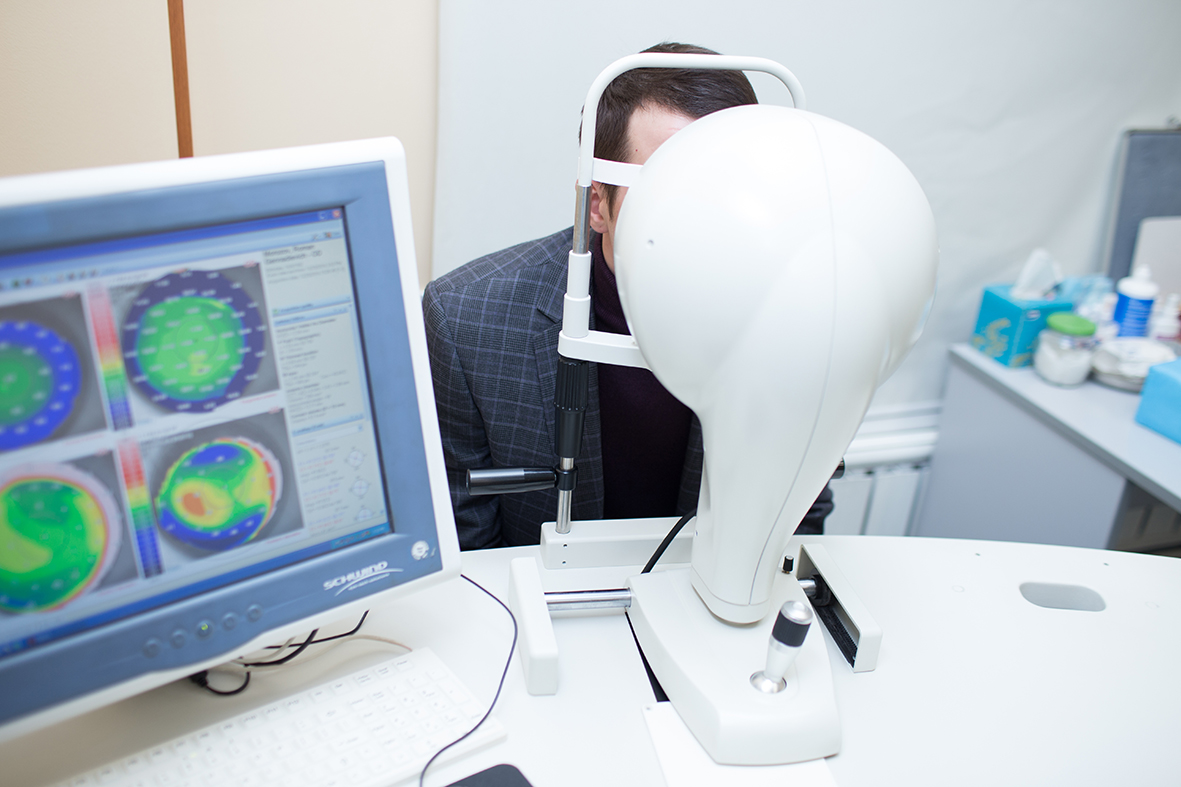

And this is the pearl of our collection - Sirius keratopograph. It allows a lot, in particular, is capable of obtaining a three-dimensional model of the cornea with the smallest details with great precision.

Corneal topography of a healthy person

Topography of a patient suffering from keratoconus. Pay attention to the sharp decrease in the thickness of the cornea in the section of corneal thickness.
I'll leave these frames here as a teaser. Sirius is clearly worthy of a separate post.
Bonus
For the keyword Geektimes, we will give you a discount of 1000 rubles for a comprehensive diagnosis. Just let his girlfriend call center when you sign up. The promotion will be valid until the end of the month (from 15.05 to 31.05) . We will be glad to see you. Let's have a normal survey)
Contacts:
"Ophthalmological clinic" Sphere "by Professor Eskina"
call center telephone: +7 (495) 480-75-84
our address: Moscow, st. Starokachalovskaya, 6
Source: https://habr.com/ru/post/374501/
All Articles