Scientists are changing laboratory mice to devices "mini-brain on a chip"
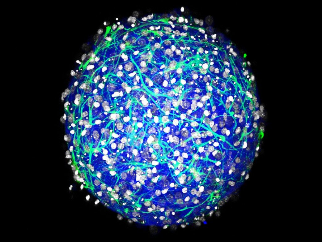
Two years ago, Diane Hoffman-Kim [Diane Hoffman-Kim] raised her first brain-ball. She started with several mouse nerve cells placed in a special Petri dish with a non-adherent surface. The cells, which had nothing to hold on to, except for each other, grew into a sphere with a diameter of less than a millimeter: a mini-brain. Bioengineers have since grown thousands of such organoids, in which neurons are active and send electrical signals. Their only negative - they are not alive. Without an independent supply of blood, they will not survive if they are not taken care of.
And then last year, one of the students, Hofman-Kim, noticed something that had not been noticed before: her globular brains spontaneously grew blood vessels.
')
This interlacing of tubules marks the beginning of the simplest circulatory system. “They can be called newbies,” says Hofman-Kim. Her mini-brains are not able to pump their blood - for this you need a heart - but this does not stop Hofman-Kim from trying to bring them closer to a state of independent life. She is working with colleagues from Brown University on connecting mini-brains to a mini-circulatory system: entire rows of balls are attached to chips connected to the motherboard with blood supply.
Over the past five years, researchers have created a large number of different micro-organs growing in Petri dishes, from small intestines to liver size Lilliputian. At the same time, progress was made in the creation of biochips: small systems the size of a USB flash drive, containing one or more layers of living cells, dotted with biosensors and microchannels for supplying liquids. These two-dimensional chips are convenient for testing, for example, the reaction of lung cells to a toxin, but too simplified to truly imitate the operation of an entire organ. And here such organoids as Hoffman-Kim's globular brains appear on the scene. For the first time, two-dimensional biochips are crossed with three-dimensional mini-organs - and together they constitute the best organ simulator for today.
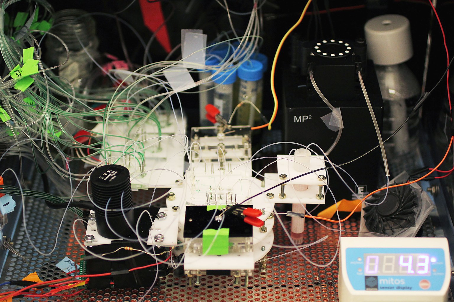
In theory, using such installations, scientists will be able to take, say, several cells of your skin, grow small versions of all your organs, and place them on a chip. Then the doctors will be able to check the effect of the drugs for the diseases you have — not on mice, but on your miniature version. “This will open a new era in personalized medicine,” says Ali Khademhosseini, a bioengineer at the Harvard Institute of Bioengineering. Viisa, who worked on the creation of mini-organs and biochips for the last ten years.
The paper, which is being prepared for publication, describes how Khademhoseini and the team created several chips that combine liver organelles and cancer cells using tubular loops. They pumped cancer medication through the system, and monitored whether it kills cancer cells, and whether liver cells survive. In this way, it is possible to dose the medication in order to maximize the anti-cancer effect without damaging the liver.
This method of testing drugs will help develop new therapies faster and cheaper. DARPA makes major investments in such research, especially focusing on treating people from the effects of nuclear and biological weapons, as this is hard to test on humans. This may mark the end of an era of drug testing on animals. Now all new drugs must be tested for animal toxicity before the developer can apply for testing in humans. This is especially important for the treatment of diseases specific to humans, since in such cases animal models do not work.
Enteroviruses each year cause 10 million infections, and for infants they are deadly. However, none of the 71 strains of the virus infects mice or rats. “If you think about it, almost everything we know about infections is based on experiments with mice,” says Carolyn Coyne, a microbiologist at the University of Pittsburgh. Therefore, Coyne has created micro-guts. A paper published last month describes how her team made human stem cells form seven different types of cells that make up the intestines. As in the case of the Hoffman-Kim mini-brains, the Coyne cells self-organize into lumps of proto-intestines, including the villi [protrusions on the mucosal surface - approx. trans.]. Some of the enteroviruses infect only certain types of cells, using them to enter the bloodstream, where they can cause maximum damage.
At the same time, mini-intestines alone were not enough to understand why the virus attacks specific cells. Coyne suspects that the case may be in the intestinal microflora . She has not yet succeeded in testing the hypothesis, since most intestinal microbes do not live in a Petri dish with its mini-intestine longer than a couple of days. But they could live longer on the chip ...
Source: https://habr.com/ru/post/373203/
All Articles