Created composite fiber, which transmits optical, electrical signals and fluid to the brain (and back)
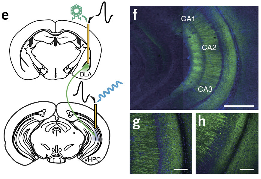
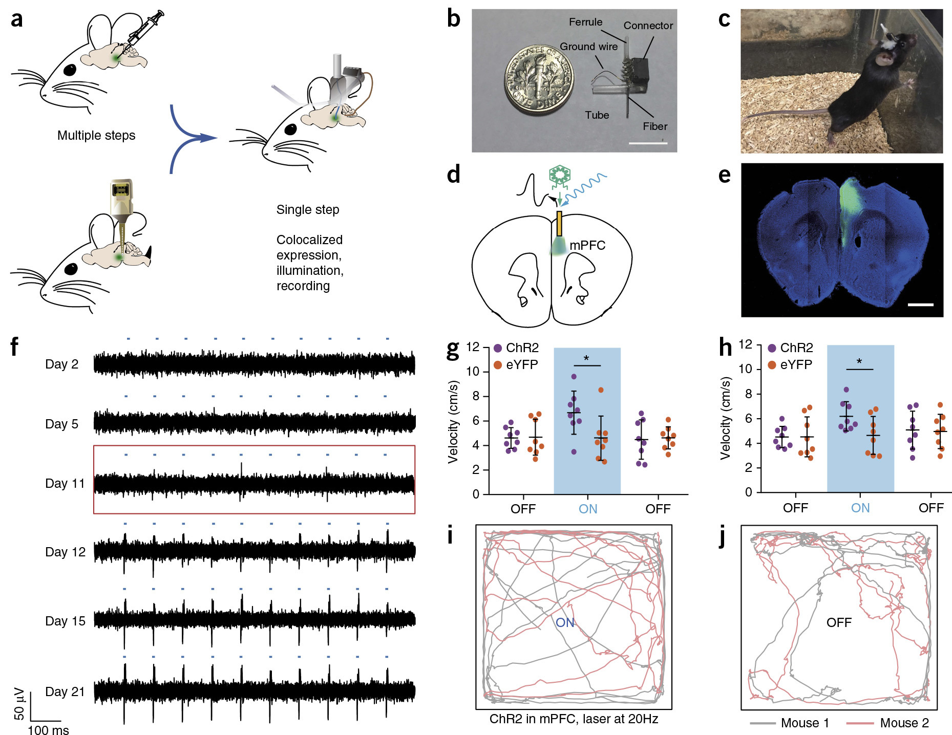
An experiment with the introduction of a solution with opsins into the mouse brain, illuminating neurons and recording their sensitivity to light. Neurons in the brain begin to react to light under the influence of opsins. The whole experiment was carried out using one electrically conductive fiber with two channels for the liquid and six electrodes.
Polina Anikeeva and colleagues at the Massachusetts Institute of Technology developed the world's first seamless flexible fiber with a thickness of less than 200 microns, which simultaneously transmits a combination of optical, electrical and chemical signals. Optical fiber transmits data in both directions - to the brain and from the brain.
The main objective of fiber in the short term - biological experiments. Actually, this fiber was invented specifically for one specific experiment. But if you improve this material and improve its biocompatibility, then it can find real application in neural interfaces.
New fibers are developed in collaboration with a large team of specialists in chemistry, biology, materials science, and other sciences. The material is designed with such a view as to best match the properties of brain tissue. Although maximum compatibility has not yet been achieved, but optics will seamlessly integrate into this fabric, retaining their properties for much longer than conventional metal fiber pins that are used now. Thus, the fiber will allow to collect more data for a longer period, and this means a significant expansion of the experimental field.
')
For example, during one experiment, scientists injected viral vectors with opsins, a group of membrane-related photosensitive proteins that were found in photosensitive cells of the retina, into the brain of a mouse. Five groups of opsins are involved in vision, the transmission of light in electrochemical signals. They are the first stage in the cascade of visual transduction. It is with the help of opsins that neurons begin to react to light.
Opsins were introduced to mice through one of two channels for fluid in the optical fiber.
Next, the researchers wanted to see how the opsins would take action. They waited some time, and then illuminated the neurons with visible light through an optical waveguide in the center of the optical fiber - and recorded the resulting activity of the neurons with six electrodes designed for specific neuron reactions.
So, all of the above was carried out using a single fiber-optic channel with a thickness of less than 200 micrometers. In order to carry out such an experiment in the past, we would have to use a number of different devices: needles for introducing viral vectors, optical fibers for illumination, and separate arrays of electrodes for signal removal. Such an experiment would be an order of magnitude more difficult if it could be done in a reliable and repeatable way at all.
Polina Anikeeva , a graduate of the St. Petersburg State Polytechnic University (2003) and the Faculty of Materials Science and Engineering at the Massachusetts Institute of Technology (2009), has been developing this material with a group of colleagues for several years - and finally it was successfully demonstrated. The scientific work was published on February 20, 2017 in the journal Nature Neuroscience (doi: 10.1038 / nn.4510). Although associate professor Anikeeva has 35 published scientific articles and 8 patents, this publication in Nature Neuroscience can be considered an important milestone in scientific career.
The small diameter of the fibers allows you to connect them simultaneously to different parts of the brain - and collect information from all departments at once. In one experiment, you can study one section of the brain, in another experiment - another section. In the past, probes with a diameter greater than 400 microns were required to read the signal from the brain using electrodes to compensate for the relatively low electrical conductivity of commercially available polymers. Now their diameter is significantly reduced. In addition, this channel can be used for additional manipulations with the medulla: illumination, fluid injection, etc. It is very convenient.
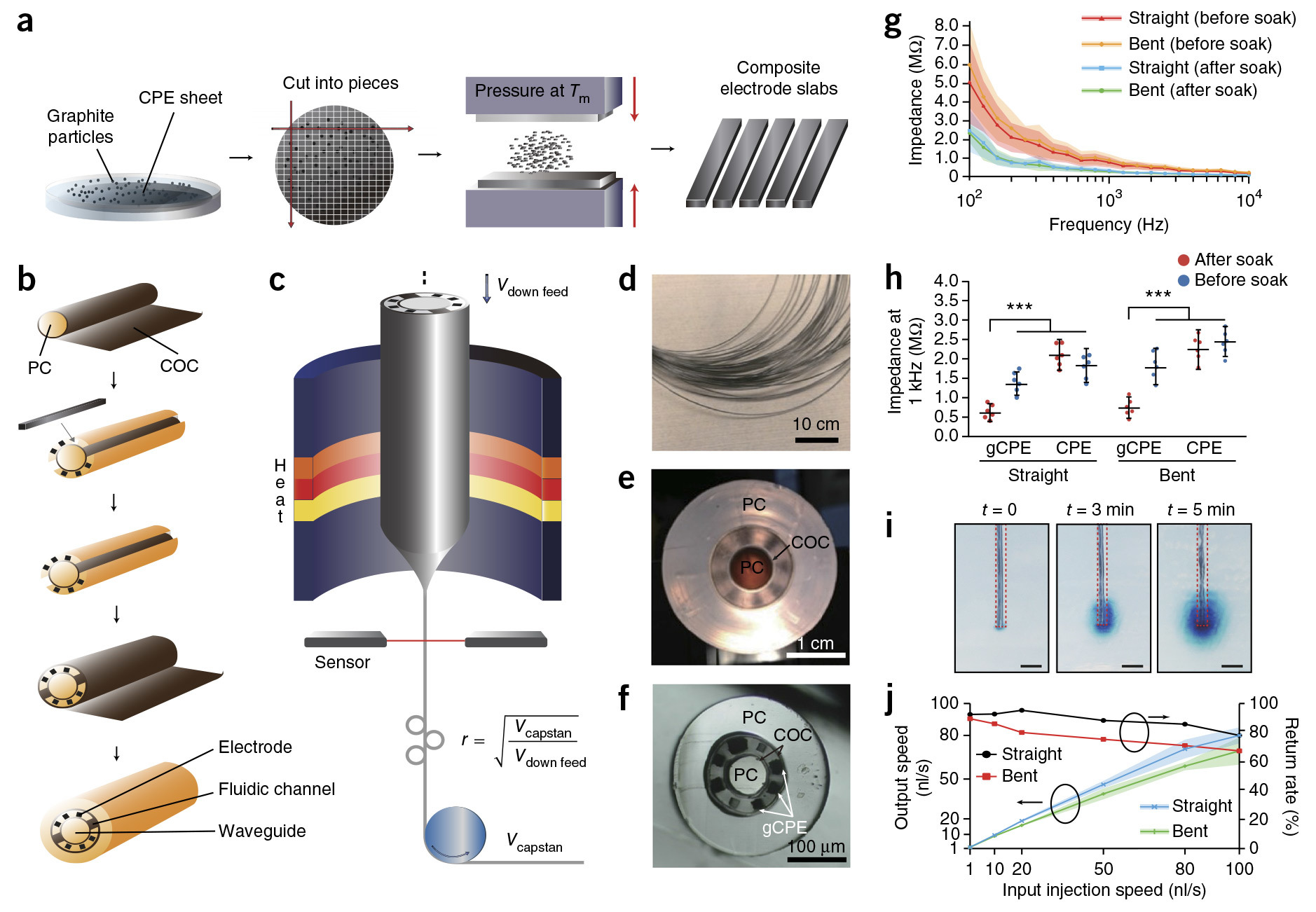
Technically, a material with such properties was able to be made by combining separate channels of composite material, which have the necessary flexibility, but at the same time conduct an electric current. They were made of conductive polyethylene doped with graphite chips. Polyethylene was formed by layers, between which sprinkled graphite, then compressed - and so on with each layer. This design allowed several times to increase the electrical conductivity of polyethylene and at the same time reduce the fiber cross section. The technical process of manufacturing a polymer composite called gCPE is shown in the illustrations.
The use of new material allowed us to finally conduct the same experiment with a long-term study of the photosensitivity of neurons after interacting with the opsins.
Previously, scientists could only make a rough estimate of how long the photosensitivity is maintained. Now they know that when using this particular agent, it lasts for about 11 days.
In general, the implanted probe continued to record the activity of neurons for more than three months, that is, the material already has quite good biocompatibility.
Source: https://habr.com/ru/post/373201/
All Articles