Neurobiological methods and cool things you can do with them: part 2
Last time, we talked about using fMRI to read thoughts and how eye tracking is associated with virtual reality. And what about the methods of creating machines, controlled by brain waves? What about technology for recognizing mental illness at the molecular level?
Electroencephalography sounds like torture technology, but the worst thing that can happen to you is a dream caused by a boring experiment. EEG is non-invasive and harmless. This method first makes people look stupid, wearing a strange construction on their heads, and then measures the electrical activity inside the head.

')
As you may be aware, neurons communicate with each other by transmitting electrical signals. When one neuron issues a signal, its activity is extremely small. But when voltage changes happen to thousands of neurons at the same time, they generate an electric field that is strong enough to pass through the skull and be found on the surface of the head. These stupid hats are used to record these signals: they contain electrodes that measure electrical activity. It is first increased 10,000 times (it is not so easy to pass through the pia mater, the skull and the scalp! The signal becomes very weak), and then transmitted to the EEG monitor, where you can watch your brain waves (or, if you prefer, sequence of stress values).
The EEG is excellent for monitoring brain activity in real time and tracking changes of a second per second (compared to him, fMRI always lags behind). But the spatial characteristics of this technology are not so good - you have to use complex algorithms to assess the exact location of the signal. There are several separate patterns of brain waves, each with its own individuality - imagine that your brain shifts gears depending on the current task. There are delta waves (usually found during deep sleep), theta waves (sleep), alpha waves (relaxation and drowsiness), beta waves (attention) and gamma waves (one of the unsolved mysteries of neuroscience; perhaps from view pictures of cats).
Watching your brain waves is an impressive accomplishment, but what about those machines that are controlled by the brain? In fact, we can come up with cool NF scenarios for using EEG: it can be used to control things with the help of consciousness (or to create brain-computer interfaces). Potential application extends from controlling a virtual helicopter in 3D to managing the limbs of robots — all with the help of thoughts. Of course, for the players, the benefits of technology are indisputable (imagine that you will not have to hold on to the joystick, and you can play with a piece of pizza in your hands), but most of all this technology will help people with disabilities.
So how do these weak electrical signals occurring inside the skull turn into movements of a roboruki? The technology is based on the idea that a certain mental activity leads to the appearance of certain EEG-signal patterns. The device will record different pictures of clumsy waves, corresponding, for example, to raising a hand, and, say, a mental pronunciation of “yes” when looking at a letter. In order to control brain activity and make these signal patterns clearly distinguishable from each other (and, thus, distinguishable for algorithms), patients (or gamers) will have to train their imagination abilities very strongly. Feedback helps: for example, every time you represent a certain movement, an arrow appears on the computer showing the direction in which you want to move. After teaching the user how to use the power of imagination, software connected with robot hands (a foot, a penis, a computer cursor) is pumped into various encephalogram drawings to recognize what you want and send commands that activate what it controls.

EEG, fMRI, and eye tracking are all very interesting, but what is really on the cutting edge of science? Spoiler: very small things. It is extremely important for us to understand the microscopic processes going on inside and between neurons, and this is what cell neuroscience is doing. You can do a lot with cells: now I work in a laboratory, where we calculate how many cells originate in the hippocampus - our memory center - in various conditions (high carbon diet, exercise, etc.). Cell behavior can be studied in vivo — in vivo, in vitro — in a flask outside the hapless living organism mentioned, and in situ — somewhere in the middle, a type like studying the brain without removing it from an already dead mouse (or, like us, dark scientists , love to say, sacrificed).
An example of an in vivo experiment is to take a mouse that is genetically modified so that the neurons of interest to us fluoresce, make a hole in its skull, and use cool technology called " two-photon microscopy " to observe fluorescent cells located relatively deep in the brain. It sounds complicated, and you can believe me, it is really complicated. In fact, your mouse is awake, but its head is fixed, and the microscope shines with high-energy light (from the blue edge of the visible spectrum) to its brain. Fluorescent cells absorb light, are excited, and emit low-energy light (green, red, yellow, etc.). The microscope catches this light through a special filter and voila - you have a beautiful picture!
Fluorescence microscopy can also be used when you have to sacrifice a mouse during an experiment. Then the brain is removed, cut into thin slices and bombarded with all sorts of chemical solutions to make its cells fluorescent. Usually, as a first step, the lobules are lowered into a solution with antibodies that connect with the cells or proteins of interest to us. In the second step, we add fluorescent antibodies that bind to the first antibodies.

I heard that you like antibodies, so I added some antibodies to your antibodies.
And then the cells become bright and shiny, and we can put them under a fluorescent microscope and enjoy the new batch of beautiful pictures!
Fluorescent Hippocampus
Cellular neuroscience has made a significant contribution to understanding the development of mental diseases and the actions necessary for their treatment. Thanks to her, we found that during depression, your hippocampus produces fewer cells than usual, and antidepressants reverse this process, stimulating the production of proteins that help neurons grow; that in some cases it is enough to increase the rate of neurogenesis (birth of new cells) to reduce anxiety and depression; that chronic stress destroys a special protein that plays an important role in ensuring communication between neurons, and that antidepressants restore its production, and mice feel better. We have learned that dopamine imbalance develops in schizophrenia, and that neuronal stem cells are not properly produced in neurons in these patients; We were also able to find new strategies for preventing Alzheimer's disease. Quite a lot of facts in a short time, so here's another beautiful picture!
And again beautiful hippocampus
Electroencephalography (EEG) and brain-computer interface
Electroencephalography sounds like torture technology, but the worst thing that can happen to you is a dream caused by a boring experiment. EEG is non-invasive and harmless. This method first makes people look stupid, wearing a strange construction on their heads, and then measures the electrical activity inside the head.
')
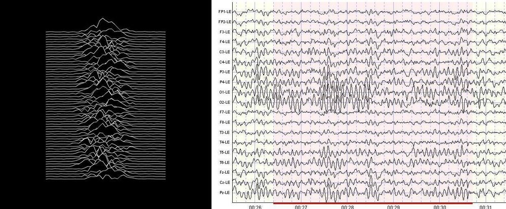
As you may be aware, neurons communicate with each other by transmitting electrical signals. When one neuron issues a signal, its activity is extremely small. But when voltage changes happen to thousands of neurons at the same time, they generate an electric field that is strong enough to pass through the skull and be found on the surface of the head. These stupid hats are used to record these signals: they contain electrodes that measure electrical activity. It is first increased 10,000 times (it is not so easy to pass through the pia mater, the skull and the scalp! The signal becomes very weak), and then transmitted to the EEG monitor, where you can watch your brain waves (or, if you prefer, sequence of stress values).
The EEG is excellent for monitoring brain activity in real time and tracking changes of a second per second (compared to him, fMRI always lags behind). But the spatial characteristics of this technology are not so good - you have to use complex algorithms to assess the exact location of the signal. There are several separate patterns of brain waves, each with its own individuality - imagine that your brain shifts gears depending on the current task. There are delta waves (usually found during deep sleep), theta waves (sleep), alpha waves (relaxation and drowsiness), beta waves (attention) and gamma waves (one of the unsolved mysteries of neuroscience; perhaps from view pictures of cats).
Watching your brain waves is an impressive accomplishment, but what about those machines that are controlled by the brain? In fact, we can come up with cool NF scenarios for using EEG: it can be used to control things with the help of consciousness (or to create brain-computer interfaces). Potential application extends from controlling a virtual helicopter in 3D to managing the limbs of robots — all with the help of thoughts. Of course, for the players, the benefits of technology are indisputable (imagine that you will not have to hold on to the joystick, and you can play with a piece of pizza in your hands), but most of all this technology will help people with disabilities.
So how do these weak electrical signals occurring inside the skull turn into movements of a roboruki? The technology is based on the idea that a certain mental activity leads to the appearance of certain EEG-signal patterns. The device will record different pictures of clumsy waves, corresponding, for example, to raising a hand, and, say, a mental pronunciation of “yes” when looking at a letter. In order to control brain activity and make these signal patterns clearly distinguishable from each other (and, thus, distinguishable for algorithms), patients (or gamers) will have to train their imagination abilities very strongly. Feedback helps: for example, every time you represent a certain movement, an arrow appears on the computer showing the direction in which you want to move. After teaching the user how to use the power of imagination, software connected with robot hands (a foot, a penis, a computer cursor) is pumped into various encephalogram drawings to recognize what you want and send commands that activate what it controls.
Cell Neurobiology and Mental Discrimination Recognition
EEG, fMRI, and eye tracking are all very interesting, but what is really on the cutting edge of science? Spoiler: very small things. It is extremely important for us to understand the microscopic processes going on inside and between neurons, and this is what cell neuroscience is doing. You can do a lot with cells: now I work in a laboratory, where we calculate how many cells originate in the hippocampus - our memory center - in various conditions (high carbon diet, exercise, etc.). Cell behavior can be studied in vivo — in vivo, in vitro — in a flask outside the hapless living organism mentioned, and in situ — somewhere in the middle, a type like studying the brain without removing it from an already dead mouse (or, like us, dark scientists , love to say, sacrificed).
An example of an in vivo experiment is to take a mouse that is genetically modified so that the neurons of interest to us fluoresce, make a hole in its skull, and use cool technology called " two-photon microscopy " to observe fluorescent cells located relatively deep in the brain. It sounds complicated, and you can believe me, it is really complicated. In fact, your mouse is awake, but its head is fixed, and the microscope shines with high-energy light (from the blue edge of the visible spectrum) to its brain. Fluorescent cells absorb light, are excited, and emit low-energy light (green, red, yellow, etc.). The microscope catches this light through a special filter and voila - you have a beautiful picture!
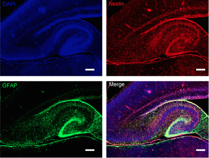
Fluorescence microscopy can also be used when you have to sacrifice a mouse during an experiment. Then the brain is removed, cut into thin slices and bombarded with all sorts of chemical solutions to make its cells fluorescent. Usually, as a first step, the lobules are lowered into a solution with antibodies that connect with the cells or proteins of interest to us. In the second step, we add fluorescent antibodies that bind to the first antibodies.
I heard that you like antibodies, so I added some antibodies to your antibodies.
And then the cells become bright and shiny, and we can put them under a fluorescent microscope and enjoy the new batch of beautiful pictures!
Fluorescent Hippocampus
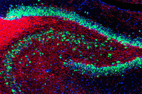
Cellular neuroscience has made a significant contribution to understanding the development of mental diseases and the actions necessary for their treatment. Thanks to her, we found that during depression, your hippocampus produces fewer cells than usual, and antidepressants reverse this process, stimulating the production of proteins that help neurons grow; that in some cases it is enough to increase the rate of neurogenesis (birth of new cells) to reduce anxiety and depression; that chronic stress destroys a special protein that plays an important role in ensuring communication between neurons, and that antidepressants restore its production, and mice feel better. We have learned that dopamine imbalance develops in schizophrenia, and that neuronal stem cells are not properly produced in neurons in these patients; We were also able to find new strategies for preventing Alzheimer's disease. Quite a lot of facts in a short time, so here's another beautiful picture!
And again beautiful hippocampus
Source: https://habr.com/ru/post/372933/
All Articles