"Flying midges" and "glassy worms" in the eyes, or where do "broken pixels" in the vitreous body come from
Lift your head and look at something evenly colored, on some light background (on snow, on the sky without the sun). If you suddenly began to swim before your eyes here are about these things:
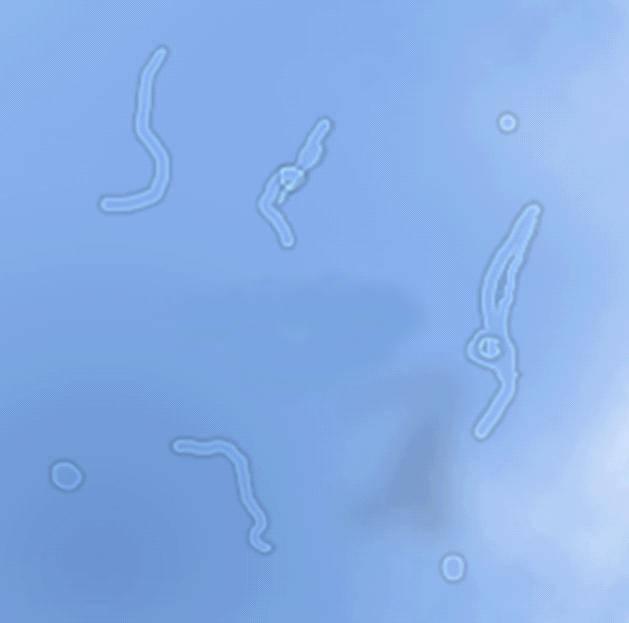
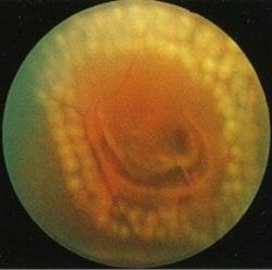
... Then get acquainted, these are “broken pixels” in your eye , formed by the vitreous body (in the figure below it is in all its glory). Many of these “glitches” appear in childhood and over the years, they multiply or gradually change. For most people, their presence is not a cause for concern, but their sudden appearance or a sharp increase is a reason for an urgent visit to an ophthalmologist. Especially if lightning before eyes, dark veil or fine “tobacco dust” are added to this.
')
But let's understand what the whole situation is and where it comes from to understand the full situation.
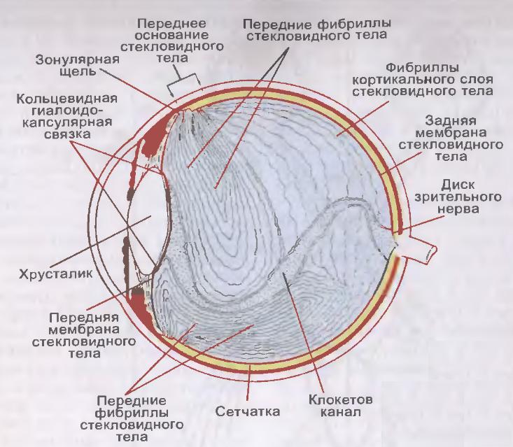
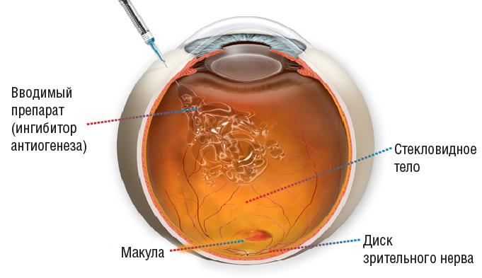
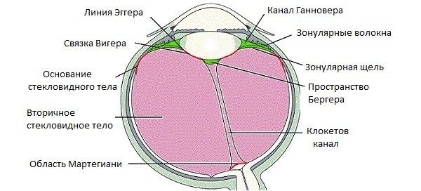
The eye is a ball, most of which is occupied by the vitreous body (as much as 2/3 volume). It is clearly seen in the diagram above - this is the space between the lens and the retina in the eye cavity. In a normal eye, the vitreous body is so transparent that when translucent eyes appear empty.
The vitreous body is a jelly-like viscous and well-stretching slurry like jelly or jelly. Only transparent. It consists of “jelly” of water, colloids and trace elements - collagen fibers resembling interwoven ropes impregnated with hyaluronic acid. Unlike the cornea, consisting of the same matrix, the density of the filaments in the vitreous is lower, so the cornea is dense and rigid (by the standards of what is in the eye), and here it is the viscous medium that awaits us.
This environment is heterogeneous, there are voids and “tanks” in it, various lacunae. You can inject a special solution into the eye that will color the vitreous and all this beauty will be visible. For example:
The vitreous body is adjacent to the back surface of the lens, for the rest of its length it comes into contact with the inner boundary membrane of the retina. From the optic nerve head to the lens through the vitreous body passes a special hyaloid channel, and the framework of the vitreous body forms a thin network of intertwined fibers of various forms of collagen protein. And the gaps are filled with liquid - this structure gives it the appearance of a gelatinous mass.
Due to the vitreous body of the eye, we have the correct spherical shape, it provides incompressibility and eye tone, it is dampened by tremors, nutrients move along the channels. But the light refractive function is very small.
If we need to deliver the medicinal substance to the deep parts of the eye, then we introduce it directly into the vitreous cavity by a micro-needle, because the eye is an organ that is rather isolated from the organism as a whole, and not everything that gets into the blood reaches the inner contents . Interferes with hematophthalmic barrier.
It happens like this:
For the most part, the translucent "ghosts" that appear in the field of view when, for example, a sharp fall or a parachute jump, lifting of gravity or against the background of complete well-being and subsequently visible when looking at bright objects carefully, are natural lacunae in the vitreous due to it by construction. They sometimes close themselves, move, or form new ones (slow, for months) by themselves.
In general, any noticeable “worms” are something in the vitreous that interferes normally with the light reaching the retina. In English literature, referred to as "floaters" -
like specks of dust on the camera's matrix. This condition is called "destruction of the vitreous body" (DST).
The condition of the presence of small single fragments in the vitreous cavity is the norm from a medical point of view.
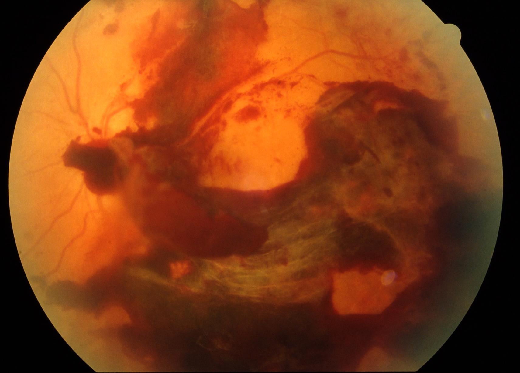
Often there is another story. But now we need a little deeper into the anatomy. In itself, the vitreous body is not attached to the retina over the main area, but simply very closely adjacent. However, in the macula (the center of the eye, the yellow spot), there are attachments near the optic nerve and at the equator of the retina, which are quite strong. If in the eye with age, with injury or the appearance of another disease of the eye and the organism as a whole, not just destruction (in general, not dangerous), but any blood cells, inflammation appears - this is a very dangerous problem. Everything that gets into such a closed cavity is absorbed for a long time, it is difficult and not always completely with a transparent effect. As a rule, dyed opacities, rough adhesions and cords, reducing visual acuity, remain. Everything is explained by relatives, I would even say, intimate contact with such an important tissue as the retina (the retina and the vitreous in the case of illness suffer simultaneously).
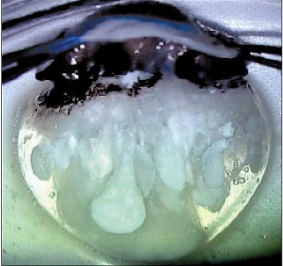
Such a terrible picture can be seen with hemorrhage into the vitreous (this is called hemophthalmus).
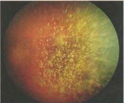
And if droplets of cholesterol accumulate in the vitreous body, it looks like a “golden rain”.
When bluntly hitting the eye, the vitreous will take on the main deformation as a result of a mechanical injury and begin to change shape. Where it is simply adjacent to the retina, it will boldly move back and return. But where there were adhesions, the vitreous body during deformation will pull the retina behind itself into the eye. From this a much more serious injury may already be formed - a rupture or detachment of the retina. And this is all the chances of losing sight or degrading the optical quality of the eye to a few percent of the norm.
Retinal tear, laser delimited
In myopic people, as a rule, the axial length of the eye is more than 24 mm (the average statistical parameter, the measurement of which tells us about the progression of myopia). The eye of the shape of a soccer ball turns into a rugby ball. At the same time, the posterior pole of the eye is stretched, but if it is not dangerous for the outer sclera (it is elastic enough), then the middle (choroid) and the inner shell (retina) do not stretch. Therefore, retinal nutrition is impaired in the posterior pole, and dystrophic zones of sprains and ruptures occur along the periphery. A large role in this just plays the vitreous body. At the point of attachment, it pulls the retina, and holes are formed.
Somewhere after 30 years, new “midges” often begin to appear, and after 40 years, hyaluronic acid is gradually lost, the transparency of the vitreous body decreases, and visualization of fibers appears.
Still later, the vitreous body generally dries out and begins to exfoliate from the retina (it simply moves away in places where there is no attachment).
Vitreous detachment is a normal sign of aging, it is caused by dilution of the vitreous body, leading to tension on the retina in places of strong attachment, which can lead to rupture of the retina. Acute detachment of the vitreous body in 15% of cases leads to retinal breaks.
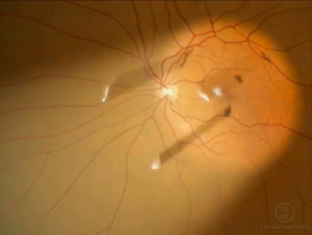
Figuratively speaking, a kind of "snot" from the gel (this is a very inaccurate description of this type of tissue, but gives a very good understanding) dangles inside the eye. Then this "lump of gel" comes off the optic nerve. As a rule, the separation zone looks like a ringlet (Weiss ring), that is, when projecting onto the retina, it turns out what patients call "spider", "big midge", "eight", "dark spot", "analemma", "circle " and so on. Kaleidoscope can change: every day a new form. If the ring of fastening has not come off, but just stretched out - hell with it, with a kaleidoscope, you can live. But if it pulled the retina and tore off a piece, then there are retinal breaks, detachments, and in general a lot of trouble.
This is how an ophthalmologist sees Weiss's ring:
Treatment of ruptures before retinal detachment occurs - laser coagulation of rupture zones in several rows along the edge so that the formed fusion sites hold the retina in place.
All this is treated with a good prognosis in the case of treatment during the first days to an ophthalmologist surgeon, with an average prognosis in the case of treatment within a month. If delamination has begun and the fluid that has entered through the gap continues to flow under the retina, exfoliating it, the problems begin to become irreversible, and the question is about preserving the vision as such, at least with some kind of optical quality.
Therefore, if you suddenly saw a lot of new "black flies", or they somehow behave strangely themselves, or something else is happening in the eye, you should immediately go to the ophthalmologist.
If, after examination, the doctor does not find damage to the retina in the fundus and only notes the presence of micro-inclusions in the vitreous, then there is no need to worry - there are no serious risks to your eyesight.
If the patient sees at the same time “garbage and midges” that bother him, but knows that there is no danger in this, the issue of dynamic observation and habituation to minor defects in the quality of vision are most often discussed. In addition, after a couple of months, these inclusions patients really cease to notice and do not focus their attention on them.
But in some cases, complaints may be due to reduced vision due to "turbidity" floating in front of the eye. Then, together with an ophthalmologist, a specialist in retina (and not just a doctor in the clinic, who know something about modern ophthalmology for quite some time), the question of treating this condition can be discussed.
There are two options: the first one involves the destruction of fragments in the vitreous body using a modern YAG laser for laser vitreolysis, the second - the surgical removal of a part of the vitreous body within the optical axis - a vitrectomy operation.
The whole trick with the problem of "midges" and "spiders" is how to find a doctor who correctly interprets changes in the eye. The difference in case of incorrect diagnosis is colossal: to distinguish the degenerative calm state of simple destruction of the vitreous body from the pathological appearance of inflammatory cells in the vitreous body, blood, the onset of retinal detachment, vascular disorders and other things.
That is, the patient faces the task of finding the expert who calms him down or notices the problem in time.
The difference in further tactics: the destruction does not need to be treated, you need to get used to it.
In clinics with an obvious picture of the DST (the usual "garbage" in the eyes without the risk of further complications), they say: "And let's appoint an absorbable therapy." And they prescribe some "placebo": emoxipin, taufon, katkhrom. "Holy water" in this case will help better if you believe in it. It is pleasant for a person, and for a couple of weeks the brain builds a map of “dead pixels” and removes them on their own (at a high level, if you start to peer, they will be visible again). If you start catching the “glitch” on purpose (as you might be doing right now), the distortion will be more noticeable. If you don’t think about them, you won’t be seen. What is required. The brain adapts well.
Another thing - to overlook the inflammation or the beginning of retinal detachment. Here, “soothing” methods cause the patient to lose a week or two, which will then lead to serious complications.
We agree that we are talking about a degenerative change in the vitreous body, and not about the other above-mentioned conditions. That is, when it is reliably known that there are no risks from a medical point of view, but something that is floating before my eyes still makes it hard to see.
Option 1. Laser vitreolysis.
Any work in a closed vitreous cavity is potentially dangerous. It must be done very precisely, since such sensitive structures as the retina (especially its central zone) and the lens are nearby.
The company Ellex has recently proposed a new minimally invasive method for treating the pathology of the vitreous body: Weiss rings, intravitreal opacities and adhesions, and degeneration of the vitreous body. This is a YAG laser system for treating the anterior and posterior parts of the eye using an aiming red diode laser. Since the structures adjacent to the vitreous body are very gentle, they invented a decrease in the total energy due to the ultra-Gaussian profile of the laser beam. The small size of the spot and low optical breakdown energy (less than 1.8 mJ in air) are performed by photodestruction of inclusions in the vitreous body.
In practice, the efficiency is 50%, because if the opacity is located close to the retina or close to the back lens capsule, or too dense, well, etc., then the application is impossible. Often, one large fragment turns into several smaller ones, they can move from the optical axis, or they can be added. In general, only after an examination by a specialist with experience in laser surgery at this facility will you be able to find out if this is your method.
Option 2. Vitrectomy.
This is a full-fledged abdominal surgery with all the ensuing consequences. That is, it does not matter whether the small “midge” is swimming or the “big spider” - the technology assumes the work of a vitreoretinal surgeon. Vitrectomy, the operation to remove the vitreous body, takes place as follows:
Three small punctures are made into the cavity of the eyeball in the projection of the plane of the ciliary body, that is, 3.5–4 mm from the limb - the border of the transparent and opaque part. The size of the puncture is measured by the geychi G (imperial unit radius). Standard 3-port seamless technology - 23 G. Now most of the operations carried out by the method of 25G - this is 0.445 millimeters. The dependence is this: the larger the G, the smaller the size of the puncture. The most gentle technique is 27G (0.361 mm). By the way, the smaller the puncture, the more expensive the cost of a set of consumables. To remove floaters perfect 27 g.
One port is supplied with a solution balanced in salt and pH, in order to maintain the tone of the eye and keep its volume during the operation, so that when the vitreous body is removed, the eye does not begin to “collapse”. The second incision is needed to shine inside this "cave", that is, the cavity inside the eye, where the vitreous body is located. Actually, the instrument is inserted into the third - with a vitreot, such a small guillotine with a pipe. It looks like a micro-meat grinder, with which the surgeon crushes the vitreous fibers and sucks them into the cavity of the tube.
At the end of the operation, a balanced solution remains in the eye, which is then replaced with intraocular fluid. Ports are removed from self-sealing joints, and a bandage is applied for a couple of hours.
In experienced hands, this operation takes 20 minutes under local anesthesia, and the result is the absence of coarse floating flakes before the eyes, but any operation with opening the eyeball has a large range of potential risks, surgery of the posterior segment of the eye doubles these risks.
In any case, I always let the patient know that this is a very serious intervention with a minimal problem from a medical point of view.
From time to time with rough opacities that reduce vision, it is necessary to perform such an operation for drivers, for pilots, etc., in the case when even a light short-term “fogging” can affect safety.
In my favorite Fedorovskiy center, such patients who are worried about trifles because of the "midge" tell a story about a patient who has come to the clinic with a complaint of "midge". He was told that it is not dangerous, it is not necessary to treat. He insisted that the doctor from the Fedorov clinic strongly discouraged the operation. He even offered to visit a psychiatrist to find the answer to the question “How to live with such a“ midge ”?”. The patient did not persuade the doctor from the Fedorov clinic for the operation and flew to look in the States for someone who would agree to operate on him. Found. After surgery, inflammation of the eye membranes occurred. The eye went blind, became small, red, and began to disturb the patient. Already in Russia, the eye had to be removed. Final: no eye - no "blacks".
How basically the body needs this structure - the vitreous body - is very difficult to judge. It seems possible without him, but the need for it in the structure of the eye is not yet fully understood. While it is intuitively clear that it is better to save, if possible.
A large number of doctors of the last century and the current one investigate the structure of the vitreous body - this is evident from the number of names by which its structures are named (channels, ligaments, etc.).
With age, the vitreous body ages, shrinks, and can cause certain problems. You need to keep an eye on your own eyes; if you experience symptoms of vision deterioration in any form or age, ask for help from specialists.
... Then get acquainted, these are “broken pixels” in your eye , formed by the vitreous body (in the figure below it is in all its glory). Many of these “glitches” appear in childhood and over the years, they multiply or gradually change. For most people, their presence is not a cause for concern, but their sudden appearance or a sharp increase is a reason for an urgent visit to an ophthalmologist. Especially if lightning before eyes, dark veil or fine “tobacco dust” are added to this.
')
But let's understand what the whole situation is and where it comes from to understand the full situation.
Where is this vitreous body
The eye is a ball, most of which is occupied by the vitreous body (as much as 2/3 volume). It is clearly seen in the diagram above - this is the space between the lens and the retina in the eye cavity. In a normal eye, the vitreous body is so transparent that when translucent eyes appear empty.
The vitreous body is a jelly-like viscous and well-stretching slurry like jelly or jelly. Only transparent. It consists of “jelly” of water, colloids and trace elements - collagen fibers resembling interwoven ropes impregnated with hyaluronic acid. Unlike the cornea, consisting of the same matrix, the density of the filaments in the vitreous is lower, so the cornea is dense and rigid (by the standards of what is in the eye), and here it is the viscous medium that awaits us.
This environment is heterogeneous, there are voids and “tanks” in it, various lacunae. You can inject a special solution into the eye that will color the vitreous and all this beauty will be visible. For example:
The vitreous body is adjacent to the back surface of the lens, for the rest of its length it comes into contact with the inner boundary membrane of the retina. From the optic nerve head to the lens through the vitreous body passes a special hyaloid channel, and the framework of the vitreous body forms a thin network of intertwined fibers of various forms of collagen protein. And the gaps are filled with liquid - this structure gives it the appearance of a gelatinous mass.
Due to the vitreous body of the eye, we have the correct spherical shape, it provides incompressibility and eye tone, it is dampened by tremors, nutrients move along the channels. But the light refractive function is very small.
If we need to deliver the medicinal substance to the deep parts of the eye, then we introduce it directly into the vitreous cavity by a micro-needle, because the eye is an organ that is rather isolated from the organism as a whole, and not everything that gets into the blood reaches the inner contents . Interferes with hematophthalmic barrier.
It happens like this:
Where are the "glass worms"
For the most part, the translucent "ghosts" that appear in the field of view when, for example, a sharp fall or a parachute jump, lifting of gravity or against the background of complete well-being and subsequently visible when looking at bright objects carefully, are natural lacunae in the vitreous due to it by construction. They sometimes close themselves, move, or form new ones (slow, for months) by themselves.
In general, any noticeable “worms” are something in the vitreous that interferes normally with the light reaching the retina. In English literature, referred to as "floaters" -
like specks of dust on the camera's matrix. This condition is called "destruction of the vitreous body" (DST).
The condition of the presence of small single fragments in the vitreous cavity is the norm from a medical point of view.
Often there is another story. But now we need a little deeper into the anatomy. In itself, the vitreous body is not attached to the retina over the main area, but simply very closely adjacent. However, in the macula (the center of the eye, the yellow spot), there are attachments near the optic nerve and at the equator of the retina, which are quite strong. If in the eye with age, with injury or the appearance of another disease of the eye and the organism as a whole, not just destruction (in general, not dangerous), but any blood cells, inflammation appears - this is a very dangerous problem. Everything that gets into such a closed cavity is absorbed for a long time, it is difficult and not always completely with a transparent effect. As a rule, dyed opacities, rough adhesions and cords, reducing visual acuity, remain. Everything is explained by relatives, I would even say, intimate contact with such an important tissue as the retina (the retina and the vitreous in the case of illness suffer simultaneously).
Such a terrible picture can be seen with hemorrhage into the vitreous (this is called hemophthalmus).
And if droplets of cholesterol accumulate in the vitreous body, it looks like a “golden rain”.
When bluntly hitting the eye, the vitreous will take on the main deformation as a result of a mechanical injury and begin to change shape. Where it is simply adjacent to the retina, it will boldly move back and return. But where there were adhesions, the vitreous body during deformation will pull the retina behind itself into the eye. From this a much more serious injury may already be formed - a rupture or detachment of the retina. And this is all the chances of losing sight or degrading the optical quality of the eye to a few percent of the norm.
Retinal tear, laser delimited
And what happens when myopia?
In myopic people, as a rule, the axial length of the eye is more than 24 mm (the average statistical parameter, the measurement of which tells us about the progression of myopia). The eye of the shape of a soccer ball turns into a rugby ball. At the same time, the posterior pole of the eye is stretched, but if it is not dangerous for the outer sclera (it is elastic enough), then the middle (choroid) and the inner shell (retina) do not stretch. Therefore, retinal nutrition is impaired in the posterior pole, and dystrophic zones of sprains and ruptures occur along the periphery. A large role in this just plays the vitreous body. At the point of attachment, it pulls the retina, and holes are formed.
What happens with age?
Somewhere after 30 years, new “midges” often begin to appear, and after 40 years, hyaluronic acid is gradually lost, the transparency of the vitreous body decreases, and visualization of fibers appears.
Still later, the vitreous body generally dries out and begins to exfoliate from the retina (it simply moves away in places where there is no attachment).
Vitreous detachment is a normal sign of aging, it is caused by dilution of the vitreous body, leading to tension on the retina in places of strong attachment, which can lead to rupture of the retina. Acute detachment of the vitreous body in 15% of cases leads to retinal breaks.
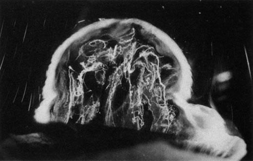
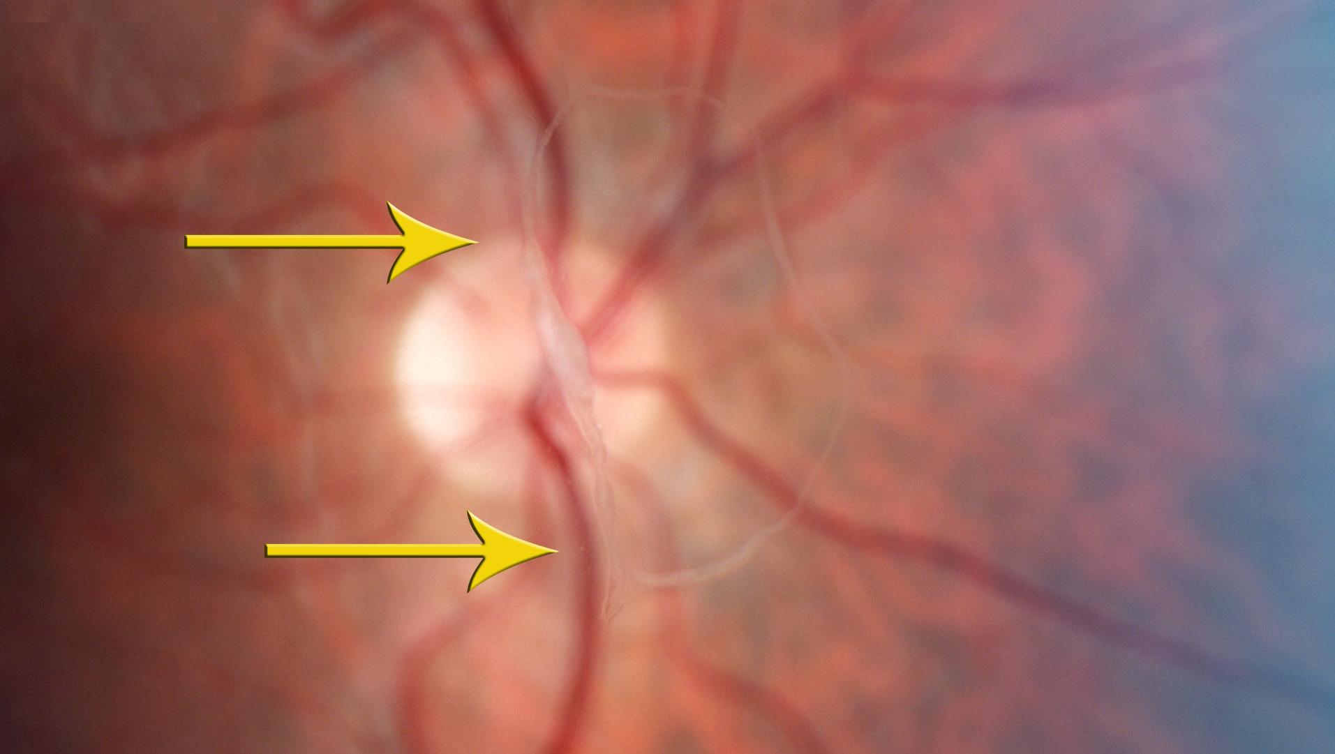
Figuratively speaking, a kind of "snot" from the gel (this is a very inaccurate description of this type of tissue, but gives a very good understanding) dangles inside the eye. Then this "lump of gel" comes off the optic nerve. As a rule, the separation zone looks like a ringlet (Weiss ring), that is, when projecting onto the retina, it turns out what patients call "spider", "big midge", "eight", "dark spot", "analemma", "circle " and so on. Kaleidoscope can change: every day a new form. If the ring of fastening has not come off, but just stretched out - hell with it, with a kaleidoscope, you can live. But if it pulled the retina and tore off a piece, then there are retinal breaks, detachments, and in general a lot of trouble.
This is how an ophthalmologist sees Weiss's ring:
Treatment of ruptures before retinal detachment occurs - laser coagulation of rupture zones in several rows along the edge so that the formed fusion sites hold the retina in place.
All this is treated with a good prognosis in the case of treatment during the first days to an ophthalmologist surgeon, with an average prognosis in the case of treatment within a month. If delamination has begun and the fluid that has entered through the gap continues to flow under the retina, exfoliating it, the problems begin to become irreversible, and the question is about preserving the vision as such, at least with some kind of optical quality.
Therefore, if you suddenly saw a lot of new "black flies", or they somehow behave strangely themselves, or something else is happening in the eye, you should immediately go to the ophthalmologist.
"Blackflies" in the eye: so what to do with them?
If, after examination, the doctor does not find damage to the retina in the fundus and only notes the presence of micro-inclusions in the vitreous, then there is no need to worry - there are no serious risks to your eyesight.
If the patient sees at the same time “garbage and midges” that bother him, but knows that there is no danger in this, the issue of dynamic observation and habituation to minor defects in the quality of vision are most often discussed. In addition, after a couple of months, these inclusions patients really cease to notice and do not focus their attention on them.
But in some cases, complaints may be due to reduced vision due to "turbidity" floating in front of the eye. Then, together with an ophthalmologist, a specialist in retina (and not just a doctor in the clinic, who know something about modern ophthalmology for quite some time), the question of treating this condition can be discussed.
There are two options: the first one involves the destruction of fragments in the vitreous body using a modern YAG laser for laser vitreolysis, the second - the surgical removal of a part of the vitreous body within the optical axis - a vitrectomy operation.
Treatment without surgery
The whole trick with the problem of "midges" and "spiders" is how to find a doctor who correctly interprets changes in the eye. The difference in case of incorrect diagnosis is colossal: to distinguish the degenerative calm state of simple destruction of the vitreous body from the pathological appearance of inflammatory cells in the vitreous body, blood, the onset of retinal detachment, vascular disorders and other things.
That is, the patient faces the task of finding the expert who calms him down or notices the problem in time.
The difference in further tactics: the destruction does not need to be treated, you need to get used to it.
In clinics with an obvious picture of the DST (the usual "garbage" in the eyes without the risk of further complications), they say: "And let's appoint an absorbable therapy." And they prescribe some "placebo": emoxipin, taufon, katkhrom. "Holy water" in this case will help better if you believe in it. It is pleasant for a person, and for a couple of weeks the brain builds a map of “dead pixels” and removes them on their own (at a high level, if you start to peer, they will be visible again). If you start catching the “glitch” on purpose (as you might be doing right now), the distortion will be more noticeable. If you don’t think about them, you won’t be seen. What is required. The brain adapts well.
Another thing - to overlook the inflammation or the beginning of retinal detachment. Here, “soothing” methods cause the patient to lose a week or two, which will then lead to serious complications.
When the operation is shown
We agree that we are talking about a degenerative change in the vitreous body, and not about the other above-mentioned conditions. That is, when it is reliably known that there are no risks from a medical point of view, but something that is floating before my eyes still makes it hard to see.
Option 1. Laser vitreolysis.
Any work in a closed vitreous cavity is potentially dangerous. It must be done very precisely, since such sensitive structures as the retina (especially its central zone) and the lens are nearby.
The company Ellex has recently proposed a new minimally invasive method for treating the pathology of the vitreous body: Weiss rings, intravitreal opacities and adhesions, and degeneration of the vitreous body. This is a YAG laser system for treating the anterior and posterior parts of the eye using an aiming red diode laser. Since the structures adjacent to the vitreous body are very gentle, they invented a decrease in the total energy due to the ultra-Gaussian profile of the laser beam. The small size of the spot and low optical breakdown energy (less than 1.8 mJ in air) are performed by photodestruction of inclusions in the vitreous body.
In practice, the efficiency is 50%, because if the opacity is located close to the retina or close to the back lens capsule, or too dense, well, etc., then the application is impossible. Often, one large fragment turns into several smaller ones, they can move from the optical axis, or they can be added. In general, only after an examination by a specialist with experience in laser surgery at this facility will you be able to find out if this is your method.
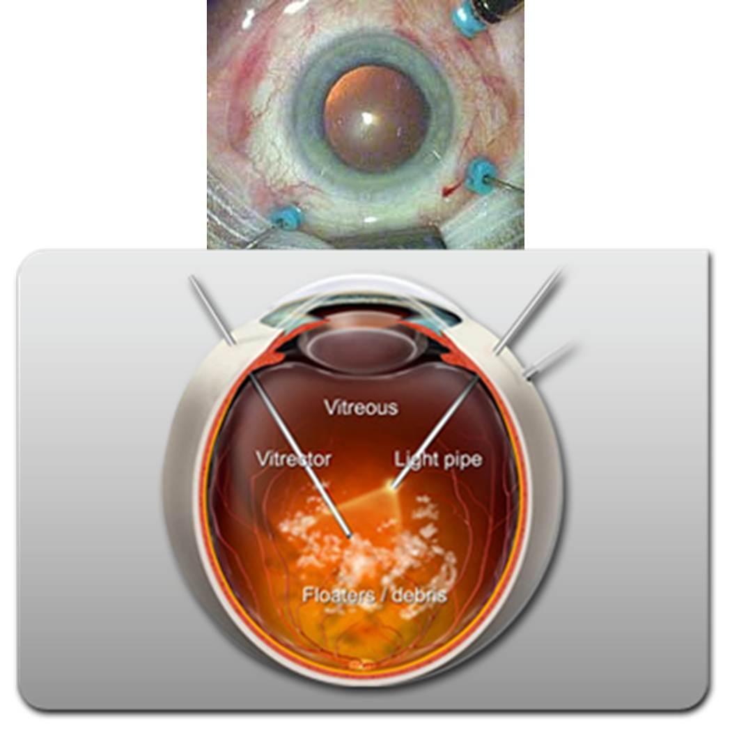
Option 2. Vitrectomy.
This is a full-fledged abdominal surgery with all the ensuing consequences. That is, it does not matter whether the small “midge” is swimming or the “big spider” - the technology assumes the work of a vitreoretinal surgeon. Vitrectomy, the operation to remove the vitreous body, takes place as follows:
Three small punctures are made into the cavity of the eyeball in the projection of the plane of the ciliary body, that is, 3.5–4 mm from the limb - the border of the transparent and opaque part. The size of the puncture is measured by the geychi G (imperial unit radius). Standard 3-port seamless technology - 23 G. Now most of the operations carried out by the method of 25G - this is 0.445 millimeters. The dependence is this: the larger the G, the smaller the size of the puncture. The most gentle technique is 27G (0.361 mm). By the way, the smaller the puncture, the more expensive the cost of a set of consumables. To remove floaters perfect 27 g.
One port is supplied with a solution balanced in salt and pH, in order to maintain the tone of the eye and keep its volume during the operation, so that when the vitreous body is removed, the eye does not begin to “collapse”. The second incision is needed to shine inside this "cave", that is, the cavity inside the eye, where the vitreous body is located. Actually, the instrument is inserted into the third - with a vitreot, such a small guillotine with a pipe. It looks like a micro-meat grinder, with which the surgeon crushes the vitreous fibers and sucks them into the cavity of the tube.
At the end of the operation, a balanced solution remains in the eye, which is then replaced with intraocular fluid. Ports are removed from self-sealing joints, and a bandage is applied for a couple of hours.
In experienced hands, this operation takes 20 minutes under local anesthesia, and the result is the absence of coarse floating flakes before the eyes, but any operation with opening the eyeball has a large range of potential risks, surgery of the posterior segment of the eye doubles these risks.
In any case, I always let the patient know that this is a very serious intervention with a minimal problem from a medical point of view.
From time to time with rough opacities that reduce vision, it is necessary to perform such an operation for drivers, for pilots, etc., in the case when even a light short-term “fogging” can affect safety.
"Scarecrow"
In my favorite Fedorovskiy center, such patients who are worried about trifles because of the "midge" tell a story about a patient who has come to the clinic with a complaint of "midge". He was told that it is not dangerous, it is not necessary to treat. He insisted that the doctor from the Fedorov clinic strongly discouraged the operation. He even offered to visit a psychiatrist to find the answer to the question “How to live with such a“ midge ”?”. The patient did not persuade the doctor from the Fedorov clinic for the operation and flew to look in the States for someone who would agree to operate on him. Found. After surgery, inflammation of the eye membranes occurred. The eye went blind, became small, red, and began to disturb the patient. Already in Russia, the eye had to be removed. Final: no eye - no "blacks".
Conclusion
How basically the body needs this structure - the vitreous body - is very difficult to judge. It seems possible without him, but the need for it in the structure of the eye is not yet fully understood. While it is intuitively clear that it is better to save, if possible.
A large number of doctors of the last century and the current one investigate the structure of the vitreous body - this is evident from the number of names by which its structures are named (channels, ligaments, etc.).
With age, the vitreous body ages, shrinks, and can cause certain problems. You need to keep an eye on your own eyes; if you experience symptoms of vision deterioration in any form or age, ask for help from specialists.
Source: https://habr.com/ru/post/370241/
All Articles