3D printing in paleontology - “resurrecting” organisms that died out millions of years ago
In the last article Digital Present and Future of Paleontology, I briefly covered the main aspects in the development of paleontology with the help of modern digital technologies. In particular, a task such as 3D modeling of fossil animals and plants was described.
But progress does not stand still, and the fact that until recently was considered fiction, in some places, is a workflow in some organizations. I mean printing with 3D printers.
I am an employee of the Darwinian Museum (Fig. 1), which, fortunately, has the ability and technology to produce such models and I would like to describe the process of building animal models from which we decided to start updating the exhibits.
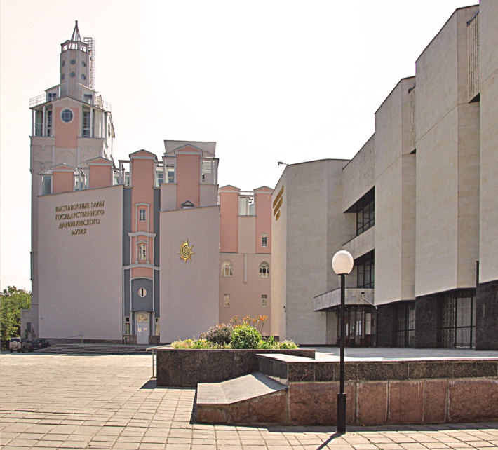
Fig. 1. Darwin Museum, Moscow, st. Vavilova, 57 (metro Akademicheskaya)
As the first trial models, we chose the fauna of the Berdess shale (Fig. 2). This unique location is located in Canada, in the Rocky Mountains and has a Middle Cambrian age (about 510 million years). At the same time, it is the richest location of Cambrian minerals in the world.
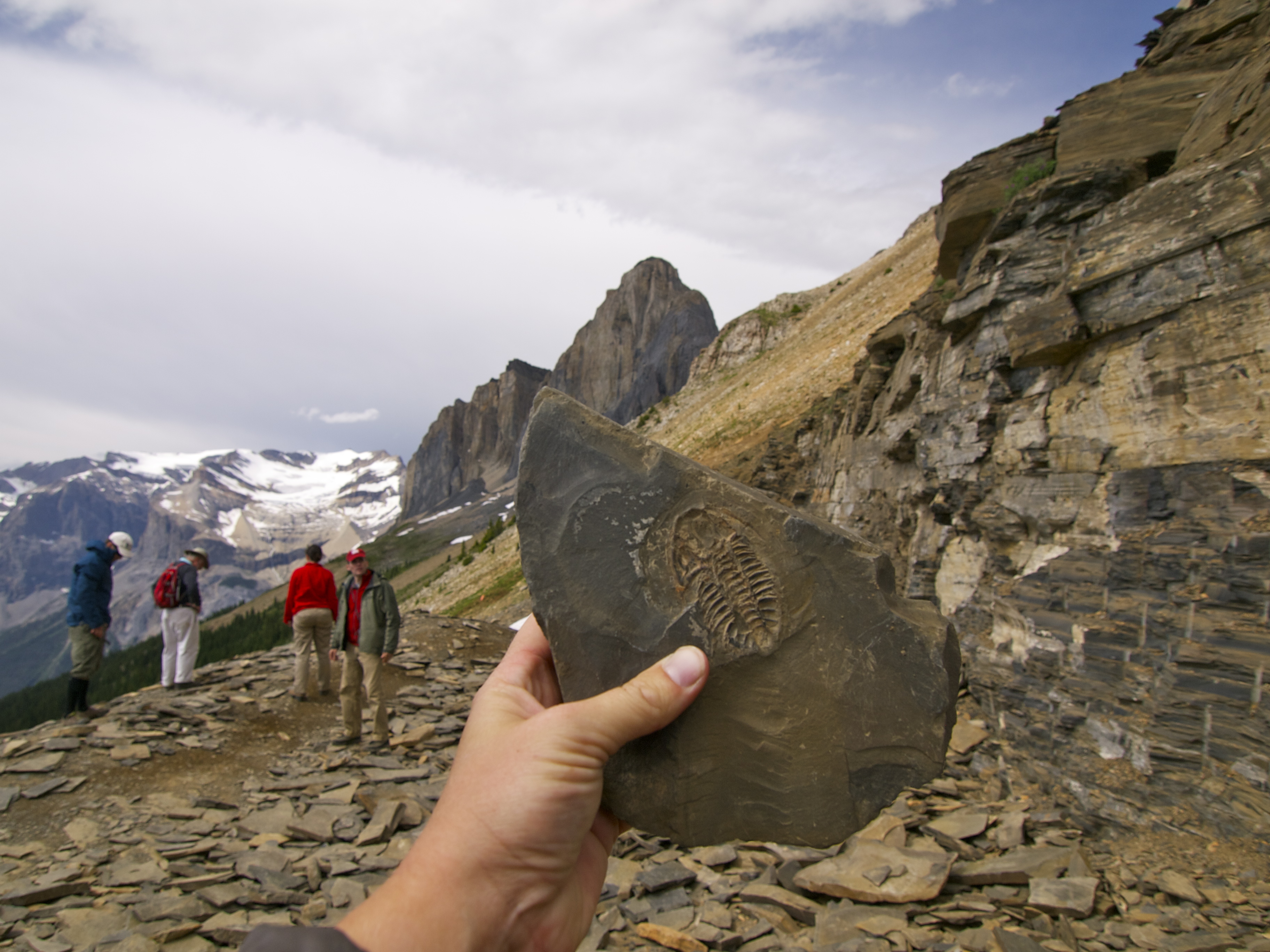
Fig. 2. Burgess Slates
A little more help on the history of the discovery of the location.
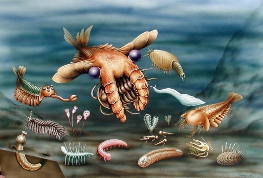
Fig. 3. Reconstruction of the animal appearance of the Burgess Shale fauna
')
We chose several of the most famous and interesting representatives of the fauna of the Burgess Shale, based on their morphology, namely, the following five types of different taxonomic groups:
Opabinia regalis
Pikaia gracilens
Anomalocaris sp.
Hallucigenia sparsa
Wiwaxia sp.
Opabinia regalis is a representative of Dinocarida class arthropods. The discoverer of opabinia, Charles Dulittl Walcott, named it in honor of the local mountain - the pass of Opabin on Mount Hungabi in the Canadian Rockies. At the moment, there are thirty known samples of opabinia, and each of them has a size in the range from 40 to 70 mm. The most remarkable feature of the abalone is its five eyes, located on the back of the head. These eyes were probably used by animals to search for food. Because of her flexible body, it is not known whether opabinia led a pelagic or benthic lifestyle.
The first reconstruction of opabiniia was presented in the work of Hutchinson (Hutchinson, 1930) and represented the opabiniia of an inverted, relatively dorsal side (Fig. 4, a).
We used as a basis the most recent reconstruction of opabinia (Fig. 4, e; Fig. 2), published a few years ago (Zhang, Briggs, 2007).
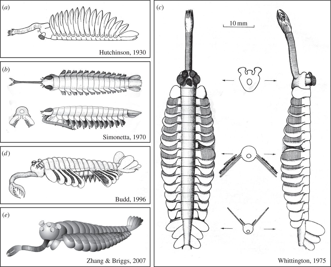
Fig. 4. The evolution of reconstructions of Opabinia regalis in the works of various authors
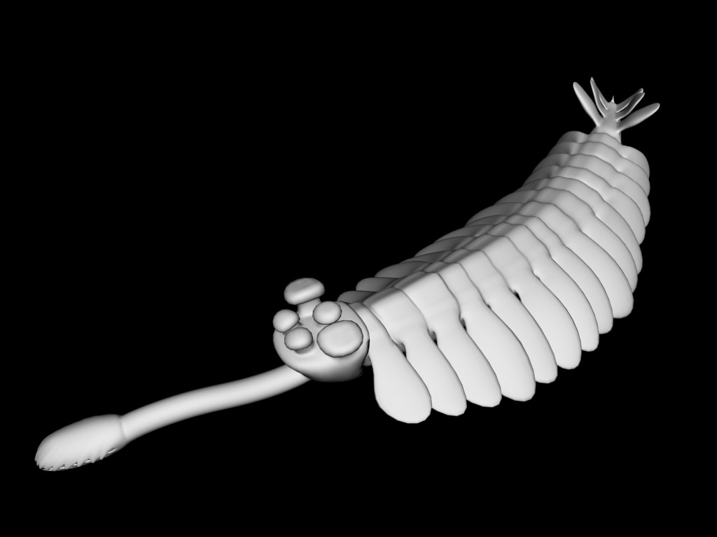
Fig. 5. Reconstruction of the Opabinia regalis, made by 3-D modeler E.Yu. Makhnev
Pikaia gracilens is a small primitive chordate animal. The body length of adult individuals was from 1.5 to 6 cm, on average, it was 4 cm. The body height was from 7 to 16% of its length, on average about 11%.
Pikaya body of elongated shape, rounded section in the front part and compressed laterally in the remaining parts. A narrow ridge resembling a fin stretched along the ventral side. Another crest, although not so high, was located on the dorsal side.
The head was very small, divided into two rounded lobes, on which there was a pair of tentacles. There is reason to believe that these tentacles were elastic, because after the death of the animal, they usually remained straight, judging by the remaining imprints. Most likely, they performed the function of organs of smell and touch. At the base of the head on its lower side there was an oral opening. No organs similar to eyes were found (Fig. 6, 7).
Behind the head on both sides of the pharynx there were 9 pairs of branchy appendages, which are interpreted by researchers as external gills. Near the bases of these appendages, small rounded spots are sometimes visible, possibly corresponding to the gill slits (Morris, Caron, 2012).
The basis was taken from the approved picaia reconstructions (Gould, 1991) and (Fig. 6).
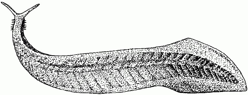
Fig. 6. Pikaya reconstruction (Gould, 1991).
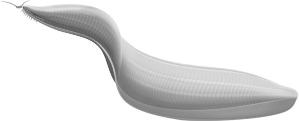
Fig. 7. A newer possible reconstruction of the appearance of Pikaia gracilens. Image from discussed article in Biological Reviews (Simon, Caron, 2012)
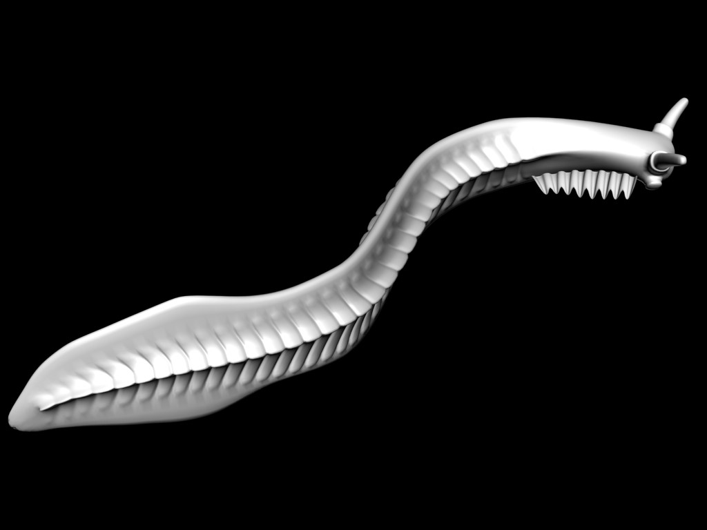
Fig. 8. Reconstruction of Pikaia gracilens, made by 3-D modeler E.Yu. Makhnev.
Anomalocaris canadensis is a type of dinocarid fossil arthropods (Dinocarida). Lived in the seas, used for swimming flexible lateral blades. Some of the largest organisms known from the Cambrian sediments: the body length could reach 60 cm and even 2 m.
The remains of the anomalokaris could not be identified for a long time - when scientists first discovered his remains, the scientists could not understand what their owner looked like. When in 1892 his long mouth appendages were found, the paleontologists decided that they belonged to some primitive crustacean. And they found the parts of the annular mouth discovered in 1911 as the remains of an ancient jellyfish. It was only in the 1980s that it became clear that the oral appendages and the plates of the mouth belonged to the same animal (Fig. 9, 10).
It was shown that the ancient anomalokarisy had excellent vision, which provided a pair of faceted eyes. Vigilance anomalokarisov provided at least 16 thousand hexagonal lenses. The presence of such complex eyes in Anomalokaris suggests that facet vision began to develop in arthropods much earlier than previously thought. It is noteworthy that, probably, the ancient anomalokaris surpassed many of its modern relatives in visual acuity. So, for comparison, the number of lenses in the eye of a fly is about 4000, and in the eye of an ant - about 100. It is noted that new results are likely to cause controversy about the evolution of the exoskeleton - in particular, how this evolution relates to the evolution of the eyes themselves (Whittington , Briggs, 1985).

Fig. 9. a - Model Anomalocaris canadensis (Whittington, Briggs, 1996), b - Model Anomalocaris in the National Dinosaur Museum, Canberra, Australia. (Wikipedia)

Fig. 10. Reconstruction of Anomalocaris canadensis, made by 3-D modeler E.Yu. Makhnev.
Hallucigenia sparsa is a type of fossil invertebrates from the Xenusia class. Discovered by English paleontologist Simon Conway-Morris in 1977. The remains were first found in the Burgess shale. Further, hallucinosis was found in the Chinese Chenchene. Externally, it looks like a worm with two rows of stilted legs and spinal spines. Were ancestral group for modern onihophore and sister group for slow moving and arthropods (Euarthropoda).
In hallucinations, an elongated head with simple eyes (Fig. 11-12), a circle of sclerotized elements in the oral cavity, and needle-shaped teeth in front of the digestive tract.

Fig. 11. a - Reconstruction (Marianne Collins in Wonderful Life Gould SJ 1991), b - Photo of the Hallucigenia sparsa holotype (Smithsonian Institute), c - Hallucigenia Sparsa reconstruction by paleoillustrator Mark Boulei, taken as the basis for modeling.

Fig. 12. Reconstruction of Hallucigenia sparsa, made by 3-D modeler E.Yu. Makhnev.
Wiwaxia is a genus of fossil soft-bodied scaly animals, known only by the fossils found for the first time in the Canadian Burges shale deposits (140 specimens were found there). Estimated time of life - from the end of the lower Cambrian to the middle Cambrian. Organisms are mainly known for multiple sclerites; The articulated specimens that were found are from 3.4 millimeters (0.13 inches) to just over 50.8 mm (2 inches) in length. The average size of the remains found is 30 mm. The appearance is somewhat reminiscent of a miniature porcupine with thorns and scales. Thorns are supposed to have served for protection. Subsequently, the fossil remains of this animal were found throughout the world, which suggests that vivaxia was widespread in the Cambrian era.
The exact taxonomic affinity of the genus is the subject of ongoing debate among paleontologists.

Fig. 13. Reconstruction of Wiwaxia

Fig. 14. Reconstruction of Wiwaxia sp., Performed by 3-D modeler E.Yu. Makhnev.
Print
On the basis of the Darwin Museum has its own Fab Lab.
Our laboratory is called CMIT ( Center for Youth Innovative Creativity ). It is here that there are several great new 3D printers. The most interesting and suitable for printing our models has become a gypsum printer brand ZPrinter450.
Further, in order not to tire with writing, I will give the process of printing in a photoset.

Fig. 15. The printing process. a) ZPrinter450 printer b) filling the bed with plaster c) printed after a couple of hours, but the object is not visible - it is necessary to clean it

Fig. 16. The cleaning process. a) vacuuming the extra plaster b) something looms)

Fig. 17. Impregnation. a) the sample is impregnated with a solution for the fortress b) the author with the finished Opabinia c) made a stand for it on the cutter
After several hours of printing and impregnation, our “soup set of the Burgess Shale fauna” is ready!

Fig. 18. a) Pikaia gracilens b) Anomalocaris sp.

Fig. 19. a) Hallucigenia sparsa b) Wiwaxia sp.
Actually, it was our "first attempt" to print fossil animal models on a 3D printer. Now we are working on color models and create a robotic model of an ancient arthropod, printed on another printer (printing plastic). But more about that in the next article.
Gould, Stephen Jay. Wonderful Life: Burgess Shale and the Nature of History, Vintage, 2000
Hutchinson, GE 1930. Restudy of some burges shale fossils. Proceedings of the US National Museum, 78: 1-11.
Conway Morris, S .; Caron, JB (2012) "Pikaia gracilens Walcott, a stem-group chordate from the Middle Cambrian of British Columbia". Biological Reviews 87: 480-512.
Whittington HB , Briggs DEG (1985) The Largest Cambrian animal, Anomalocaris, Burgess Shale, British Columbia. Philos T RoySoc B 390: 569 - 609
Zhang, X., Briggs, DEG (2007). The Cambrian Burgess Shale. Lethaia 40, 161-173.
Materials wikipedia.org
But progress does not stand still, and the fact that until recently was considered fiction, in some places, is a workflow in some organizations. I mean printing with 3D printers.
I am an employee of the Darwinian Museum (Fig. 1), which, fortunately, has the ability and technology to produce such models and I would like to describe the process of building animal models from which we decided to start updating the exhibits.
Fig. 1. Darwin Museum, Moscow, st. Vavilova, 57 (metro Akademicheskaya)
Preamble
As the first trial models, we chose the fauna of the Berdess shale (Fig. 2). This unique location is located in Canada, in the Rocky Mountains and has a Middle Cambrian age (about 510 million years). At the same time, it is the richest location of Cambrian minerals in the world.
Fig. 2. Burgess Slates
A little more help on the history of the discovery of the location.
Fossil remains in the Burgess shale were first discovered by paleontologist Charles Doolittle Walcott in 1909 at the end of his field season. In 1910 he returned there with his son and began excavations near the area, called Fossil Ridge. He returned to the excavations every year until 1924 - by the time he was 74 years old, he had discovered more than 65,000 samples. Walcott was busy describing these samples until his death in 1927. Walcott's mistake was that he tried to place all the fossil remains in taxons known by that time, and many of them looked like curiosities. Only in 1962 there was a fundamental revision of the classification of the Burgess remnants, which was conducted by Alberto Simonetta. This led to a new explosion of interest in the locality, as it turned out that many open organisms are something new, previously unknown. The number of palaeontological remains found in the formation is so large that the term “Burges Shale fauna” has come into use.
Analysis of the fossil shale Burgess, made by Whittington and his colleagues in the 1970s, formed the basis of Gould's book The Amazing Life, which opened the Cambrian explosion to the general public (Fig. 3).
Arthropods are most common among fossil Burges shale, but many of them are unusual and difficult to classify.
Fig. 3. Reconstruction of the animal appearance of the Burgess Shale fauna
')
Types and models
We chose several of the most famous and interesting representatives of the fauna of the Burgess Shale, based on their morphology, namely, the following five types of different taxonomic groups:
Opabinia regalis
Pikaia gracilens
Anomalocaris sp.
Hallucigenia sparsa
Wiwaxia sp.
Opabinia regalis is a representative of Dinocarida class arthropods. The discoverer of opabinia, Charles Dulittl Walcott, named it in honor of the local mountain - the pass of Opabin on Mount Hungabi in the Canadian Rockies. At the moment, there are thirty known samples of opabinia, and each of them has a size in the range from 40 to 70 mm. The most remarkable feature of the abalone is its five eyes, located on the back of the head. These eyes were probably used by animals to search for food. Because of her flexible body, it is not known whether opabinia led a pelagic or benthic lifestyle.
The first reconstruction of opabiniia was presented in the work of Hutchinson (Hutchinson, 1930) and represented the opabiniia of an inverted, relatively dorsal side (Fig. 4, a).
We used as a basis the most recent reconstruction of opabinia (Fig. 4, e; Fig. 2), published a few years ago (Zhang, Briggs, 2007).
Fig. 4. The evolution of reconstructions of Opabinia regalis in the works of various authors
Fig. 5. Reconstruction of the Opabinia regalis, made by 3-D modeler E.Yu. Makhnev
Pikaia gracilens is a small primitive chordate animal. The body length of adult individuals was from 1.5 to 6 cm, on average, it was 4 cm. The body height was from 7 to 16% of its length, on average about 11%.
Pikaya body of elongated shape, rounded section in the front part and compressed laterally in the remaining parts. A narrow ridge resembling a fin stretched along the ventral side. Another crest, although not so high, was located on the dorsal side.
The head was very small, divided into two rounded lobes, on which there was a pair of tentacles. There is reason to believe that these tentacles were elastic, because after the death of the animal, they usually remained straight, judging by the remaining imprints. Most likely, they performed the function of organs of smell and touch. At the base of the head on its lower side there was an oral opening. No organs similar to eyes were found (Fig. 6, 7).
Behind the head on both sides of the pharynx there were 9 pairs of branchy appendages, which are interpreted by researchers as external gills. Near the bases of these appendages, small rounded spots are sometimes visible, possibly corresponding to the gill slits (Morris, Caron, 2012).
The basis was taken from the approved picaia reconstructions (Gould, 1991) and (Fig. 6).
Fig. 6. Pikaya reconstruction (Gould, 1991).
Fig. 7. A newer possible reconstruction of the appearance of Pikaia gracilens. Image from discussed article in Biological Reviews (Simon, Caron, 2012)
Fig. 8. Reconstruction of Pikaia gracilens, made by 3-D modeler E.Yu. Makhnev.
Anomalocaris canadensis is a type of dinocarid fossil arthropods (Dinocarida). Lived in the seas, used for swimming flexible lateral blades. Some of the largest organisms known from the Cambrian sediments: the body length could reach 60 cm and even 2 m.
The remains of the anomalokaris could not be identified for a long time - when scientists first discovered his remains, the scientists could not understand what their owner looked like. When in 1892 his long mouth appendages were found, the paleontologists decided that they belonged to some primitive crustacean. And they found the parts of the annular mouth discovered in 1911 as the remains of an ancient jellyfish. It was only in the 1980s that it became clear that the oral appendages and the plates of the mouth belonged to the same animal (Fig. 9, 10).
It was shown that the ancient anomalokarisy had excellent vision, which provided a pair of faceted eyes. Vigilance anomalokarisov provided at least 16 thousand hexagonal lenses. The presence of such complex eyes in Anomalokaris suggests that facet vision began to develop in arthropods much earlier than previously thought. It is noteworthy that, probably, the ancient anomalokaris surpassed many of its modern relatives in visual acuity. So, for comparison, the number of lenses in the eye of a fly is about 4000, and in the eye of an ant - about 100. It is noted that new results are likely to cause controversy about the evolution of the exoskeleton - in particular, how this evolution relates to the evolution of the eyes themselves (Whittington , Briggs, 1985).
Fig. 9. a - Model Anomalocaris canadensis (Whittington, Briggs, 1996), b - Model Anomalocaris in the National Dinosaur Museum, Canberra, Australia. (Wikipedia)
Fig. 10. Reconstruction of Anomalocaris canadensis, made by 3-D modeler E.Yu. Makhnev.
Hallucigenia sparsa is a type of fossil invertebrates from the Xenusia class. Discovered by English paleontologist Simon Conway-Morris in 1977. The remains were first found in the Burgess shale. Further, hallucinosis was found in the Chinese Chenchene. Externally, it looks like a worm with two rows of stilted legs and spinal spines. Were ancestral group for modern onihophore and sister group for slow moving and arthropods (Euarthropoda).
In hallucinations, an elongated head with simple eyes (Fig. 11-12), a circle of sclerotized elements in the oral cavity, and needle-shaped teeth in front of the digestive tract.
Fig. 11. a - Reconstruction (Marianne Collins in Wonderful Life Gould SJ 1991), b - Photo of the Hallucigenia sparsa holotype (Smithsonian Institute), c - Hallucigenia Sparsa reconstruction by paleoillustrator Mark Boulei, taken as the basis for modeling.
Fig. 12. Reconstruction of Hallucigenia sparsa, made by 3-D modeler E.Yu. Makhnev.
Wiwaxia is a genus of fossil soft-bodied scaly animals, known only by the fossils found for the first time in the Canadian Burges shale deposits (140 specimens were found there). Estimated time of life - from the end of the lower Cambrian to the middle Cambrian. Organisms are mainly known for multiple sclerites; The articulated specimens that were found are from 3.4 millimeters (0.13 inches) to just over 50.8 mm (2 inches) in length. The average size of the remains found is 30 mm. The appearance is somewhat reminiscent of a miniature porcupine with thorns and scales. Thorns are supposed to have served for protection. Subsequently, the fossil remains of this animal were found throughout the world, which suggests that vivaxia was widespread in the Cambrian era.
The exact taxonomic affinity of the genus is the subject of ongoing debate among paleontologists.
Fig. 13. Reconstruction of Wiwaxia
Fig. 14. Reconstruction of Wiwaxia sp., Performed by 3-D modeler E.Yu. Makhnev.
On the basis of the Darwin Museum has its own Fab Lab.
Fab Lab (Fab Lab) is a production and educational workshop where you can make almost everything. The main task of the fablab is to help innovators to introduce their products from ideas to prototyping, searching for marketing solutions and creating innovative products.
Our laboratory is called CMIT ( Center for Youth Innovative Creativity ). It is here that there are several great new 3D printers. The most interesting and suitable for printing our models has become a gypsum printer brand ZPrinter450.
Further, in order not to tire with writing, I will give the process of printing in a photoset.
Fig. 15. The printing process. a) ZPrinter450 printer b) filling the bed with plaster c) printed after a couple of hours, but the object is not visible - it is necessary to clean it
Fig. 16. The cleaning process. a) vacuuming the extra plaster b) something looms)
Fig. 17. Impregnation. a) the sample is impregnated with a solution for the fortress b) the author with the finished Opabinia c) made a stand for it on the cutter
After several hours of printing and impregnation, our “soup set of the Burgess Shale fauna” is ready!
Fig. 18. a) Pikaia gracilens b) Anomalocaris sp.
Fig. 19. a) Hallucigenia sparsa b) Wiwaxia sp.
Actually, it was our "first attempt" to print fossil animal models on a 3D printer. Now we are working on color models and create a robotic model of an ancient arthropod, printed on another printer (printing plastic). But more about that in the next article.
Literature
Gould, Stephen Jay. Wonderful Life: Burgess Shale and the Nature of History, Vintage, 2000
Hutchinson, GE 1930. Restudy of some burges shale fossils. Proceedings of the US National Museum, 78: 1-11.
Conway Morris, S .; Caron, JB (2012) "Pikaia gracilens Walcott, a stem-group chordate from the Middle Cambrian of British Columbia". Biological Reviews 87: 480-512.
Whittington HB , Briggs DEG (1985) The Largest Cambrian animal, Anomalocaris, Burgess Shale, British Columbia. Philos T RoySoc B 390: 569 - 609
Zhang, X., Briggs, DEG (2007). The Cambrian Burgess Shale. Lethaia 40, 161-173.
Materials wikipedia.org
Source: https://habr.com/ru/post/368419/
All Articles