Ultrasonic technology allows you to monitor the patient's heart in real time

Cardiologist Bijoy Khandheria has been treating the hearts of his patients for over 30 years, listening to the sounds made by this body and looking at the images obtained through ultrasound. However, the doctor is not satisfied with the current state of affairs, because the ultrasound does not give the whole picture. “We use the signal to display the image layer by layer, almost like a butcher uses a knife, and then in our head we put the picture into a single whole. This process always implies some assumptions, ”says the doctor.
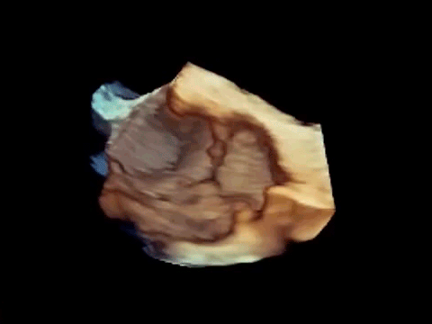
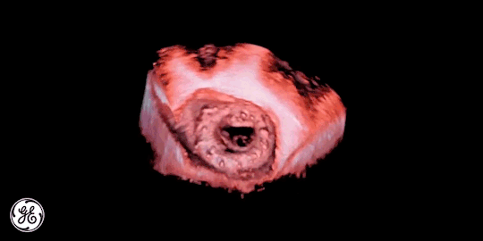
Now the situation may change as Dr. Khandheria and colleagues from Aurora St. Luke's Medical Center has created a new software platform. This platform, using the ultrasound system, shows doctors the patient's heart in 4D - three dimensions plus time. In other words, doctors see the work of a person’s heart in real time.
')
The developers claim that the platform created by the efforts of GE Healthcare specialists builds a picture so precisely that it becomes possible to even see the blood twist around the blood clots in the arteries.
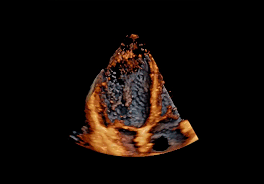
The ultrasound system sends high-frequency acoustic waves into the body, and then captures the reflection using a specific algorithm to build a “picture” visible to a person.
Usually it all depends on the equipment that generates acoustic signals. But the main thing here is the software platform, and the algorithms that allow you to create high-definition images showing all the necessary features of the structure of the heart muscle of a particular patient.
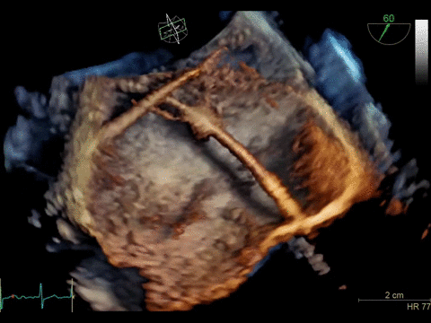
The software platform itself is called cSound. The image is based on the data analyzed by the system, with the construction of the "pixel-by-pixel" picture. The amount of data passing through the software is quite large. In just one second, the software receives as much information as one DVD holds, analyzing it all in real time. Naturally, an ordinary PC can not cope with such a load, so you have to use the capabilities of the supercomputer (which one is not reported).
The development of cSound was carried out with an eye on another ultrasound system from GE, which is used to construct an image of the fetus of a pregnant woman. Here is a slightly different scheme for analyzing data and building a picture, but the principle of operation is general.
Source: https://habr.com/ru/post/367697/
All Articles