“Laser can help diagnose cancer”, or How does a laser fluorescence hyperspectral microscope work?
Not so long ago, our press service prepared material on the studies of the Leading scientist, a member of the Corr. RAS, d.ph. - m. Khazanov Efim Arkadyevich. An interview with a leading scientist was published on a variety of resources, some of them can be found by following the links: Laser and Professor Khazanov will help diagnose and treat cancer : "soon we will learn to treat cancer cells with a laser . " It was about an innovative method of diagnosis and treatment of cancer with the help of a laser-plasma proton accelerator. In order to fully dive into the essence of the research, we asked the lead researcher Konstantin Yushkov to tell how the equipment itself is used for the research process and the creation of a prototype.
Under the cat you will find a lot of information about the project, equipment, uniqueness of the study. We have prepared a photo report that describes a system of hyperspectral analysis and laser research of microscopic preparations and reveals its significance.

Laboratory "Physical methods of acousto-optic and laser equipment for the tasks of diagnosis and therapy of oncological diseases"
Efim Khazanov, Institute of Applied Physics, Russian Academy of Sciences, is developing methods for diagnosing and treating cancer using laser technology and creating a component base for a laser-plasma proton accelerator based on the new laboratory at NITU MISiS, under the direction of Dr. Sc. one of whose applications is cancer therapy.
')
The research team of the laboratory united the efforts of leading specialists in the field of acoustooptics and image processing systems (NITU MISiS), laser physics (IAP RAS, Nizhny Novgorod), oncology (ESC RAMS, Biological Faculty of Moscow State University) crystal physics (Tver State University, Tver). As part of the project, a unique complex of research equipment was created, allowing to conduct research in the field of biomedical optics.
The main goal of the laboratory is the creation of new acousto-optic systems for hyperspectral laser fluorescence diagnostics of oncological diseases and acousto-optic devices for femtosecond laser complexes of hadron therapy of oncological diseases. This project represents a unique combination of modern developments in the field of optics and laser physics with applications in the biomedical field (oncology). The urgency of the tasks is due to the focus on the creation of key components of a new generation of hadron therapy devices for oncological diseases based on compact laser sources and high-energy charged particle accelerators. The project also solves the problem of formulating new diagnostic criteria for the differential diagnosis and determination of human cancer malignancy.
The results that were achieved by the laboratory team:
1. An experimental prototype of an acousto-optic hyperspectral system was created with the possibility of spatial filtering and contouring of images.
2. A laser photocathode driver was created for electron injection with an acousto-optic control system with ultrashort laser pulses.
3. Research in the field of acoustics and optics of anisotropic media made it possible to determine the optimal configurations of wide-aperture and quasicollinear acousto-optic filters based on paratellurite single crystals.
4. Hyperspectral studies of the fluorescent radiation of human tumor tissues of the thyroid gland were carried out.
5. Developed methods for the formation of arbitrary spectral transmission functions of acousto-optic filters.
System of hyperspectral analysis and laser research of microscopic preparations
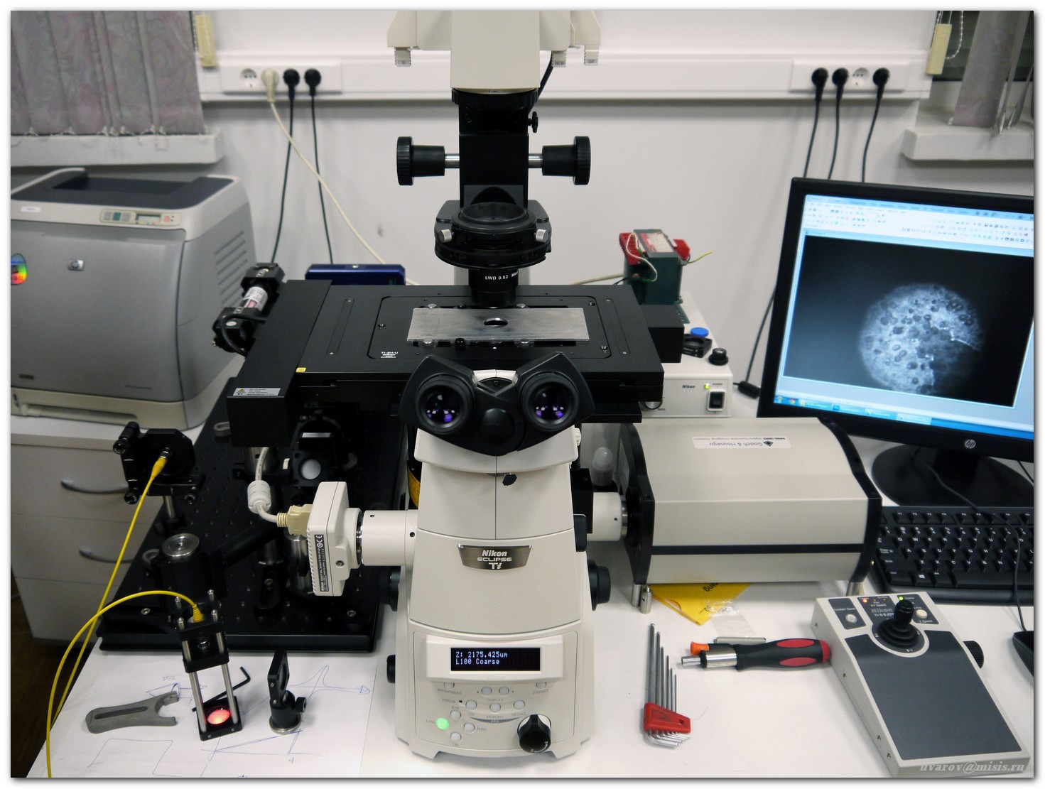
The system is based on a Nikon Ti-E inverted microscope with a set of lenses from 4x to 100x
 Visual inspection is carried out through the eyepiece or using a standard color camera.
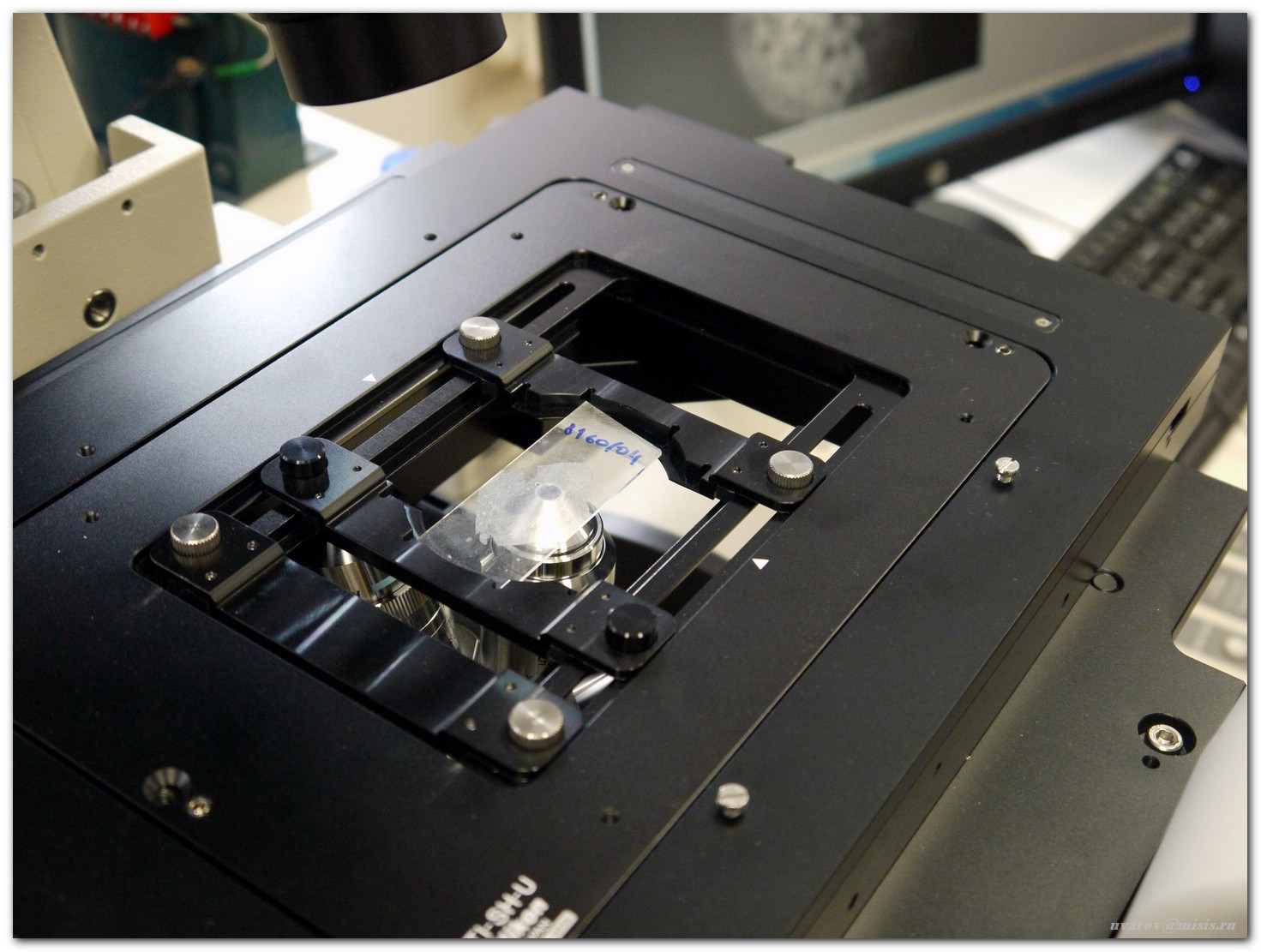 The studied samples are preparations of cytological smears and histological sections of human tumors on a high-precision motorized microscope XY-table
 Standard microscopic examination method is sample white light transmission (halogen lamp, standard microscope condenser)
 The red laser radiation is directed to the collimator, which allows for single-mode fiber to transmit light to the object
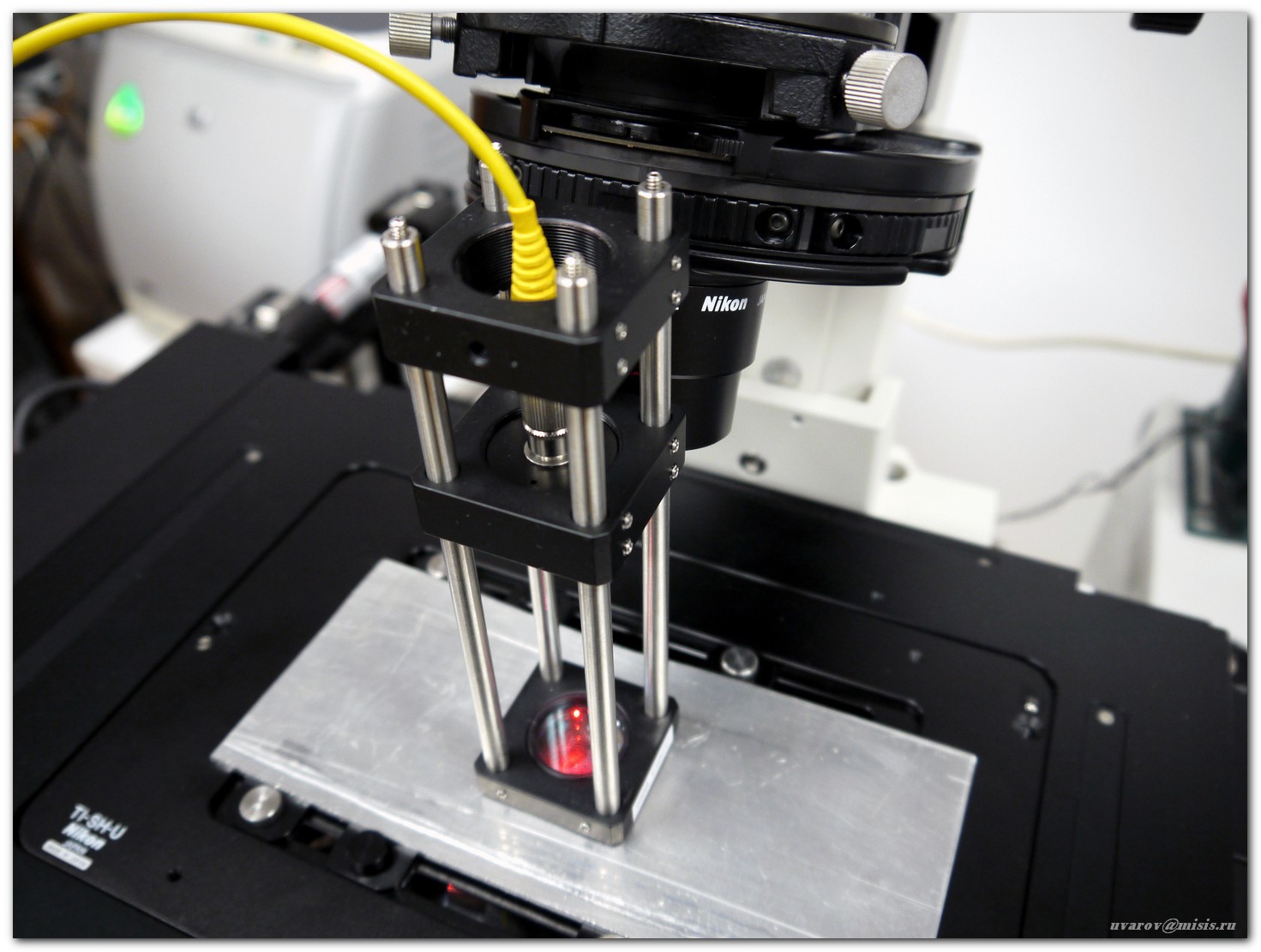 The laser illuminator of the preparation consists of a free end of the optical fiber and a lens with a focal length of 100 mm. After the lens, an almost flat front of the light wave is formed, which allows to investigate the phase structure of objects
 An enlarged image of the object is formed in the side port of the microscope, which is subjected to further processing and an acousto-optic filter.
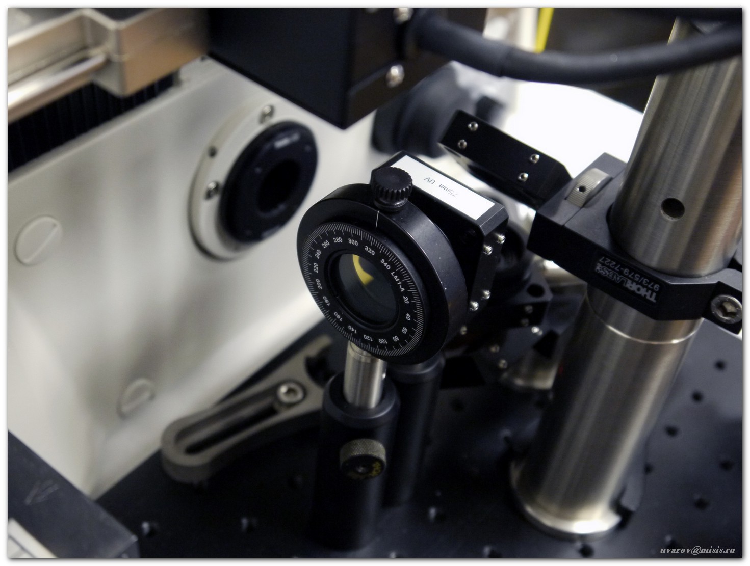 An auxiliary optical system of mirrors, aperture and polarizer forms an image at the input of an acousto-optic filter
 Acousto-optic filter, diffracted image transfer system and highly sensitive cooled CCD camera process and detect hyperspectral images
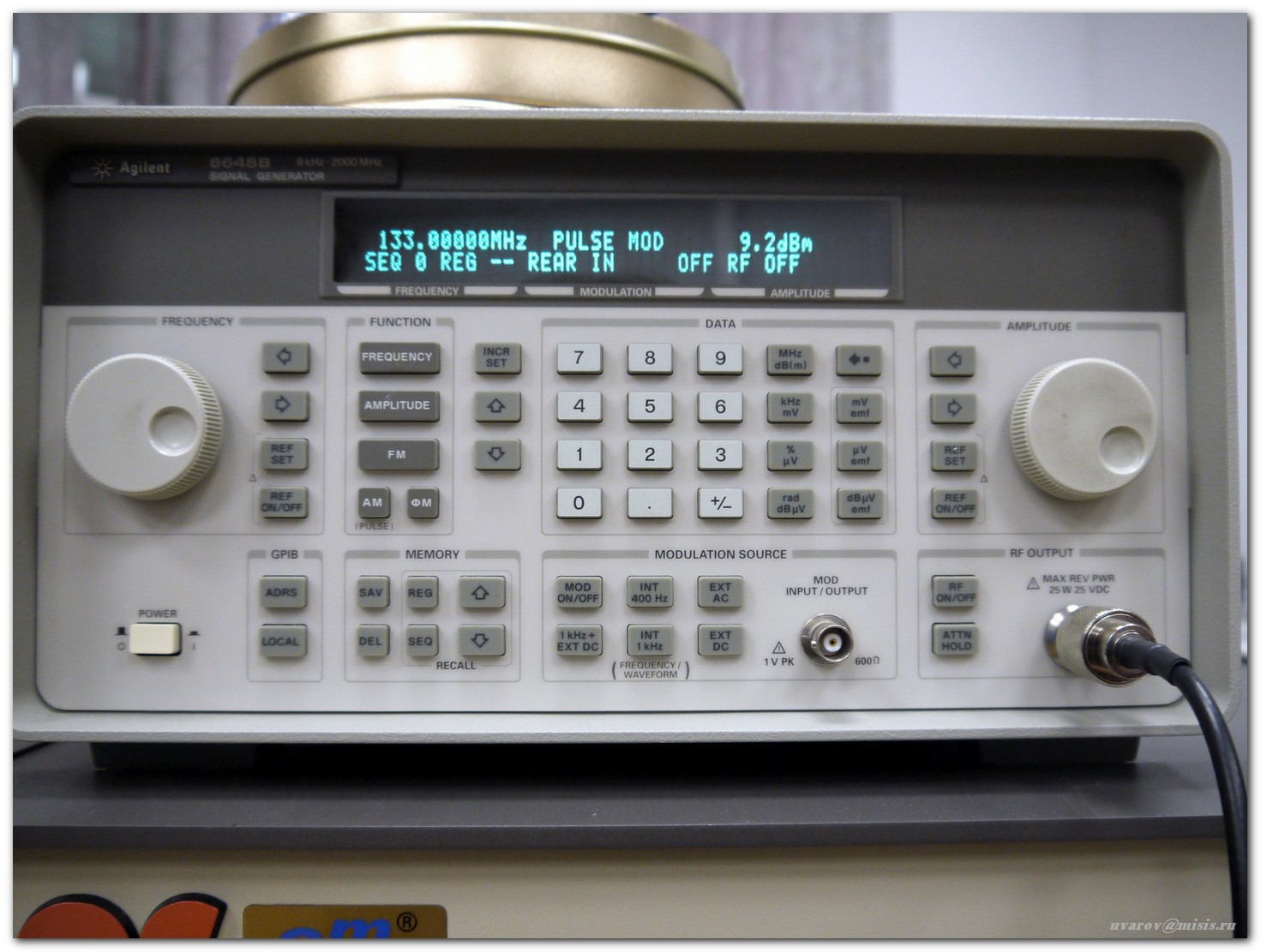 The parameters of an acousto-optic filter are controlled using a digital high-frequency generator
 Fragment of stained histological section of human follicular adenoma. Lens magnification 40x. Left: color image; in the center: hyperspectral image obtained using an acousto-optic system; right: contour image obtained using an acousto-optic system with laser illumination.
 Fluorescent studies of tumor tissue samples are carried out using a laser system of epifluorescent illumination. There are two lasers on the optical table: a green Nd: YAG laser with frequency doubling by a wavelength of 532 nm and a blue Ar laser at a wavelength of 488 nm.
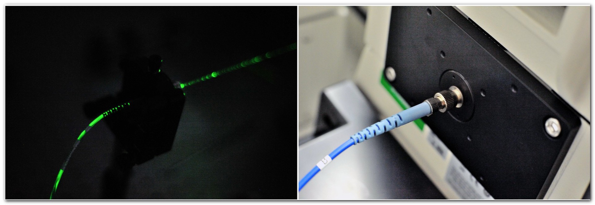 Laser radiation through a collimator is inserted into a polarization-preserving single-mode optical fiber (left), which is connected to the back port of the microscope via a special adapter (right)
 In the microscope, special beam-splitting cubes with high-contrast light filters (dichroic mirror + stopband) are used to direct the laser radiation to the object and filter the fluorescence signal.
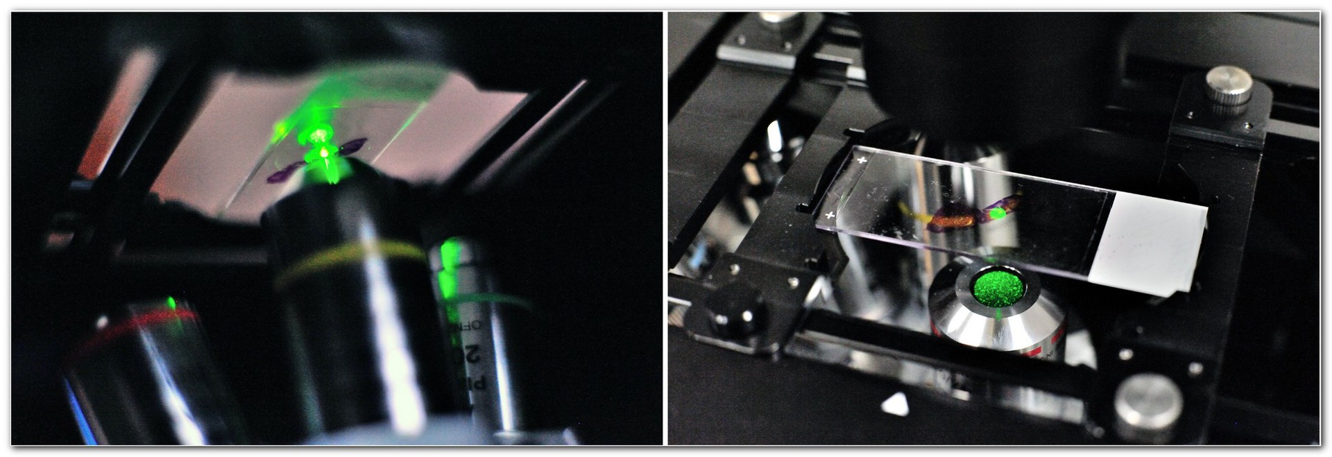 Laser illumination hits the object - a glass slide with a tissue preparation applied on it - through the same lens that builds the image on the detector
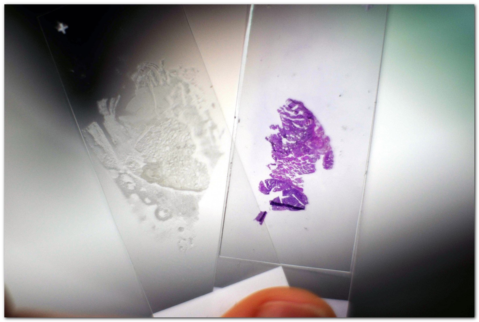 The objects of research are both stained and unstained histological sections of remote tumors of the human thyroid gland.
 Various cases are investigated: benign, malignant, and tumors of undetermined malignant potential. Comparison with reference samples of normal tissue is also performed.
Under the cat you will find a lot of information about the project, equipment, uniqueness of the study. We have prepared a photo report that describes a system of hyperspectral analysis and laser research of microscopic preparations and reveals its significance.
Laboratory "Physical methods of acousto-optic and laser equipment for the tasks of diagnosis and therapy of oncological diseases"
Efim Khazanov, Institute of Applied Physics, Russian Academy of Sciences, is developing methods for diagnosing and treating cancer using laser technology and creating a component base for a laser-plasma proton accelerator based on the new laboratory at NITU MISiS, under the direction of Dr. Sc. one of whose applications is cancer therapy.
')
The research team of the laboratory united the efforts of leading specialists in the field of acoustooptics and image processing systems (NITU MISiS), laser physics (IAP RAS, Nizhny Novgorod), oncology (ESC RAMS, Biological Faculty of Moscow State University) crystal physics (Tver State University, Tver). As part of the project, a unique complex of research equipment was created, allowing to conduct research in the field of biomedical optics.
The main goal of the laboratory is the creation of new acousto-optic systems for hyperspectral laser fluorescence diagnostics of oncological diseases and acousto-optic devices for femtosecond laser complexes of hadron therapy of oncological diseases. This project represents a unique combination of modern developments in the field of optics and laser physics with applications in the biomedical field (oncology). The urgency of the tasks is due to the focus on the creation of key components of a new generation of hadron therapy devices for oncological diseases based on compact laser sources and high-energy charged particle accelerators. The project also solves the problem of formulating new diagnostic criteria for the differential diagnosis and determination of human cancer malignancy.
The results that were achieved by the laboratory team:
1. An experimental prototype of an acousto-optic hyperspectral system was created with the possibility of spatial filtering and contouring of images.
2. A laser photocathode driver was created for electron injection with an acousto-optic control system with ultrashort laser pulses.
3. Research in the field of acoustics and optics of anisotropic media made it possible to determine the optimal configurations of wide-aperture and quasicollinear acousto-optic filters based on paratellurite single crystals.
4. Hyperspectral studies of the fluorescent radiation of human tumor tissues of the thyroid gland were carried out.
5. Developed methods for the formation of arbitrary spectral transmission functions of acousto-optic filters.
Equipment:
• Femtosecond laser Femtosource Synergy (Femtolasers, Austria).
• Femtosecond autocorrelator Femtometer (Femtolasers, Austria).
• Amplifier regenerative RAP-2000 (Avesta-project, Russia).
• Diagnostics system of ultrashort laser pulses MIIPSBox-640 (Biophotonic Solitions, USA).
• N8241A arbitrary waveform generator (Agilent, USA).
• The arbitrary waveform generator 33622 (Keysight, USA).
• Optical spectrum analyzer 86142B (Agilent, USA).
• 488 nm argon laser (Melles Griot, USA).
• Solid state laser 532 nm (Laser Quantum, UK).
• RF amplifiers (Amplifier Research, USA).
• FSH3 radio spectrum analyzer (Rhode & Schwarz, Germany).
• Radio power meter NRT-Z14 (Rhode & Schwarz, Germany).
• X-ray diffractometer (Rigaku, Japan).
• Automated installation for cutting crystals and plates APD2 (Logitech, UK).
• Grinding and polishing optical machine OLP 200 / C (AKM, Germany).
• OptoTL-60 interferometer (Optical Technology Laboratory, Russia).
• Ultrasonic washing line (Selecta, Spain).
• Vacuum spray unit (Torr, USA)
• Installation of thermocompression welding (Kulicke & Soffa, USA).
• Vector network analyzer E5061A (Agilent, USA).
• Medical biology microscope Ti-E (Nikon, Japan).
• Hyperspectral system HSi-300 (Gooch & Housego, USA).
• Cooled Alta U32 CCD Camera (Apogee Instruments, USA).
• Precision optical tables (Thorlabs, USA).
• Optical station (Thorlabs, USA).
System of hyperspectral analysis and laser research of microscopic preparations
 | Konstantin Yushkov, Leading Researcher |
Hyperspectral analysis consists in obtaining and an array of images of the object under study at different wavelengths and the subsequent study of the spectral features of various image fragments. The fundamentals of hyperspectral analysis technology, also called spectrozonal imaging, were developed for solving problems in astrophysics, space research, and remote sensing of the Earth. In recent years, these methods have been introduced in biomedical research, in particular in the diagnosis of cancer. The amount of information about an object obtained by hyperspectral analysis significantly exceeds color images: the spectral resolution of modern hyperspectral systems is several hundred lines in the visible spectrum, while a color (RGB) camera highlights only three broad spectral ranges (red, green and blue). One of the physical instruments that allow the imaging spectrometer to be implemented is tunable acousto-optic filters.
The system is based on a Nikon Ti-E inverted microscope with a set of lenses from 4x to 100x
Visual inspection is carried out through the eyepiece or using a standard color camera. The studied samples are preparations of cytological smears and histological sections of human tumors on a high-precision motorized microscope XY-table Standard microscopic examination method is sample white light transmission (halogen lamp, standard microscope condenser)The object image observed in this case contains information about the spectral transmission curves of the sample, which, however, is lost when registering with a conventional CCD camera. In addition, when observing an object in white light, only amplitude modulation can be seen, that is, the difference between more and less transparent image fragments. Phase modulation due to variations in the thickness of the sample or its refractive index can be visualized only in coherent light and with the help of special spatial filtration systems. One of the classes of such systems is acousto-optic image filters, the indisputable advantage of which is adaptability, that is, the ability to change the characteristics during the experiment, depending on the task.
The red laser radiation is directed to the collimator, which allows for single-mode fiber to transmit light to the object The laser illuminator of the preparation consists of a free end of the optical fiber and a lens with a focal length of 100 mm. After the lens, an almost flat front of the light wave is formed, which allows to investigate the phase structure of objects An enlarged image of the object is formed in the side port of the microscope, which is subjected to further processing and an acousto-optic filter. An auxiliary optical system of mirrors, aperture and polarizer forms an image at the input of an acousto-optic filter Acousto-optic filter, diffracted image transfer system and highly sensitive cooled CCD camera process and detect hyperspectral imagesAcousto-optic filter is a key element of this system and allows image processing in two fundamentally different modes:
• under white light hyperspectral analysis is performed, that is, sequential selection of different wavelengths from the spectrum of radiation transmitted by an object and registration of an array of spectral images (the so-called “hyperspectral cube”);
• with laser illumination, adaptive spatial filtering of images is performed, which allows for analog processing such as contouring (underlining intensity gradients) and phase visualization (that is, observing object optical density variations that are not recorded when illuminated with white light).
To increase the sensitivity, low-noise black-and-white cooled CCD cameras are used as detectors, and the wavelength of the saved image, that is, its color, is determined by the acousto-optical filter setting. When post-processing from a hyperspectral array of images, a color image can be restored or a false color color scheme can be used to enhance the visual contrast of the details.
The parameters of an acousto-optic filter are controlled using a digital high-frequency generator Fragment of stained histological section of human follicular adenoma. Lens magnification 40x. Left: color image; in the center: hyperspectral image obtained using an acousto-optic system; right: contour image obtained using an acousto-optic system with laser illumination. Fluorescent studies of tumor tissue samples are carried out using a laser system of epifluorescent illumination. There are two lasers on the optical table: a green Nd: YAG laser with frequency doubling by a wavelength of 532 nm and a blue Ar laser at a wavelength of 488 nm. Laser radiation through a collimator is inserted into a polarization-preserving single-mode optical fiber (left), which is connected to the back port of the microscope via a special adapter (right) In the microscope, special beam-splitting cubes with high-contrast light filters (dichroic mirror + stopband) are used to direct the laser radiation to the object and filter the fluorescence signal. Laser illumination hits the object - a glass slide with a tissue preparation applied on it - through the same lens that builds the image on the detector The objects of research are both stained and unstained histological sections of remote tumors of the human thyroid gland. Various cases are investigated: benign, malignant, and tumors of undetermined malignant potential. Comparison with reference samples of normal tissue is also performed.Comparison of images obtained by various methods: a normal color image, hyperspectral black and white images, a fluorescent image - allows you to study in detail the characteristics of various tissues and get additional information about their structure. The study of fluorescence spectra allows us to observe some features of the tissues that are not detected with conventional light microscopy.
The combination of two different physical methods of image processing in one optical scheme: hyperspectral analysis and spatial filtration is the main advantage of the system developed in NITU "MISiS" for the study of histological sections of tumor tissues. This equipment opens up new possibilities for visualization of intracellular structures and the development of new diagnostic criteria in oncology. In the future, the system will allow to implement additional imaging modes of the object and integrate new modules, such as the femtosecond excitation system of multiphoton fluorescence.
Source: https://habr.com/ru/post/366313/
All Articles