A blood test: from a light microscope to hematology analyzers
A general blood test is the most common diagnostic test prescribed by the patient by the doctor. Over the past decade, the technology of this routine, but very informative research has made a tremendous breakthrough - it has become automatic. To help the doctor of laboratory diagnostics, whose instrument was a conventional light microscope, came high-tech automatic hematological analyzers.
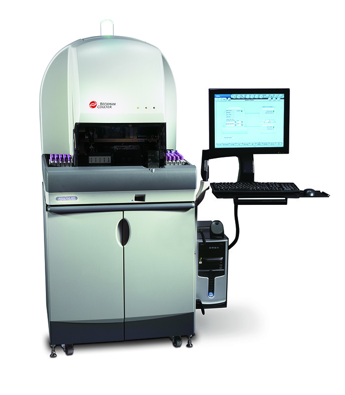
In this post we will tell you exactly what is happening inside the “smart machine” that sees through our blood and why it should be believed. We will consider the physics of processes on the example of the hematology analyzer UniCel DxH800 of the world brand Beckman Coulter. It is on this equipment that the studies ordered in the service of laboratory diagnostics LAB4U.RU are performed . But in order to understand the technology of automatic blood analysis, we will understand what the laboratory doctors saw under the microscope and how they interpreted this information.
Blood test parameters
So, the blood contains three types of cells:
- white blood cells that provide immune protection;
- platelets responsible for blood clotting;
- red blood cells transporting oxygen and carbon dioxide.
These cells are in the blood in very specific quantities. They are determined by the age of a person and his state of health. Depending on the conditions in which the body is located, the bone marrow produces as many cells as the body needs. Therefore, knowing the number of a certain type of blood cells and their shape, size and other qualitative characteristics, one can confidently judge the state and current needs of the body. It is these key parameters - the number of cells of each type, their appearance and qualitative characteristics - constitute the overall clinical blood analysis.
')
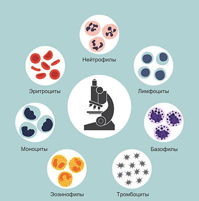
When conducting a complete blood count, red blood cells, platelets, and white blood cells are counted. With leukocytes more difficult: there are several types, and each species performs its function. There are 5 different types of leukocytes:
- neutrophils, mainly neutralizing bacteria;
- eosinophils, neutralizing antigen-antibody immune complexes;
- basophils involved in allergic reactions;
- monocytes are major macrophages and utilizers;
- lymphocytes that provide general and local immunity.
In turn, neutrophils according to the degree of maturity are divided into:
- bandker,
- segmented,
- myelocytes
- metamyelocytes.
The percentage of each type of leukocyte in their total volume is called a leukocyte formula, which has an important diagnostic value. For example, the more pronounced the bacterial inflammatory process, the more neutrophils in the leukocyte formula. The presence of neutrophils of different degrees of maturity indicates the severity of the bacterial infection. The sharper the process, the more blood in the stab neutrophils. The appearance of metamyelocytes and myelocytes in the blood indicates an extremely serious bacterial infection. An increase in lymphocytes is characteristic of viral diseases, and in allergic reactions an increase in eosinophils.
In addition to quantitative indicators, cell morphology is extremely important. The change in their usual shape and size also indicates the presence of certain pathological processes in the body.
An important and most well-known indicator is the amount of hemoglobin in the blood — a complex protein that provides oxygen to the tissues and removes carbon dioxide. The concentration of hemoglobin in the blood is the main indicator in the diagnosis of anemia.
Another important parameter is the erythrocyte sedimentation rate (ESR). In inflammatory processes in erythrocytes, the property appears to stick together with each other, forming small clots. Possessing a larger mass, stuck together erythrocytes under the action of gravity settle faster than single cells. The change in their sedimentation rate in mm / h is a simple indicator of inflammatory processes in the body.
As it was: scarifier, test tubes and microscope
Blood sampling
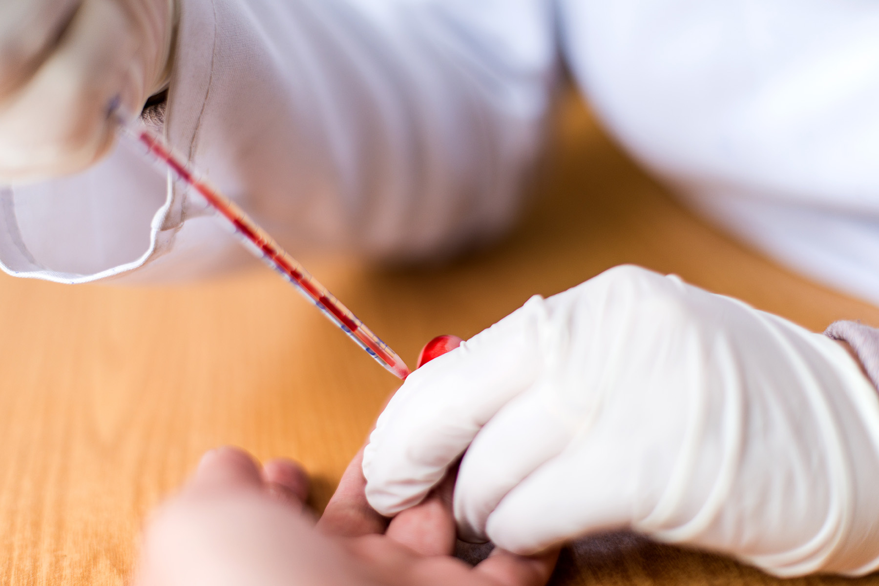
Let us recall how blood was donated earlier: a painful puncture of the pads by a scarifier, endless glass tubes into which precious drops of squeezed blood were collected. As a laboratory assistant, I used one glass to pass over another, where there was a drop of blood, scratching the number on the glass with a simple pencil. And endless test tubes with different liquids. Now it seems to be some kind of alchemy.
Blood was taken precisely from the ring finger, for which there were quite serious reasons: the anatomy of this finger is such that its injury gives minimal risk of sepsis in the event of a wound infection. Blood sampling from a vein was considered far more dangerous. Therefore, the analysis of venous blood was not routine, but was prescribed by necessity, and mainly in hospitals.
It should be noted that already at the stage of the fence began significant errors. For example, different thickness of the skin gives a different depth of injection, along with blood, tissue fluid gets into the test tube - hence the change in blood concentration, in addition, with pressure on the finger, blood cells could be destroyed.
Remember the row of tubes where the blood collected from the finger was placed? For counting different cells, we really needed different tubes. For erythrocytes - with saline, for leukocytes - with a solution of acetic acid, where erythrocytes were dissolved, to determine hemoglobin - with a solution of hydrochloric acid. A separate capillary was to determine the ESR. And at the last stage a smear was made on the glass for the subsequent calculation of the leukocyte formula.
Blood test under the microscope
To count the cells under a microscope in laboratory practice, a special optical device was used, proposed as early as the 19th century by a Russian doctor, by whose name this device was called the Goryaev camera. She allowed to determine the number of cells in a given microvolume of the liquid and was a thick glass slide with a rectangular recess (camera). A microscopic grid was applied to it. Top camera Goryaeva covered with a thin cover glass.
This grid consisted of 225 large squares, 25 of which were divided into 16 small squares. The erythrocytes were counted in small striated squares located along the Goryaev’s camera diagonal. Moreover, there was a certain rule for counting cells, which lie on the border of a square. The calculation of the number of red blood cells per liter of blood was carried out according to the formula, based on the dilution of blood and the number of squares in the grid. After mathematical abbreviations, it was enough to multiply the number of cells in the chamber by 10 to the 12th power and enter it into the analysis form.
Leukocytes were counted here, but already used large grid squares, since leukocytes are a thousand times larger than erythrocytes. After counting the leukocytes, their number was multiplied by 10 to the 9th degree and added to the blank. For an experienced lab technician, cell counting took an average of 3-5 minutes.
Platelet counting methods in the Goryaev chamber were very laborious due to the small size of this type of cells. They had to estimate their number only on the basis of a stained blood smear, and the process itself was also very laborious. Therefore, as a rule, the number of platelets was calculated only by special request of the doctor.
Leukocyte formula , that is, the percentage of leukocytes of each type in their total number could be determined only by a doctor according to the results of a study of blood smears on glasses.

Visually identifying various types of leukocytes in the field of view according to the shape of their nucleus, the doctor considered the cells of each species and their total number. Having counted 100 in aggregate, he received the required percentage of each type of cell. To simplify the counting, special counters were used with separate keys for each type of cells.
It is noteworthy that such an important parameter as hemoglobin was determined by a laboratory technician visually (!) By the color of hemolyzed blood in a test tube with hydrochloric acid. The method was based on the conversion of hemoglobin to brown hematin hydrochloride, the color intensity of which is proportional to the hemoglobin content. The resulting solution of hematin hydrochloride was diluted with water to the color of a standard corresponding to a known concentration of hemoglobin. In general, the last century
How it became: vacuum containers and hematology analyzers
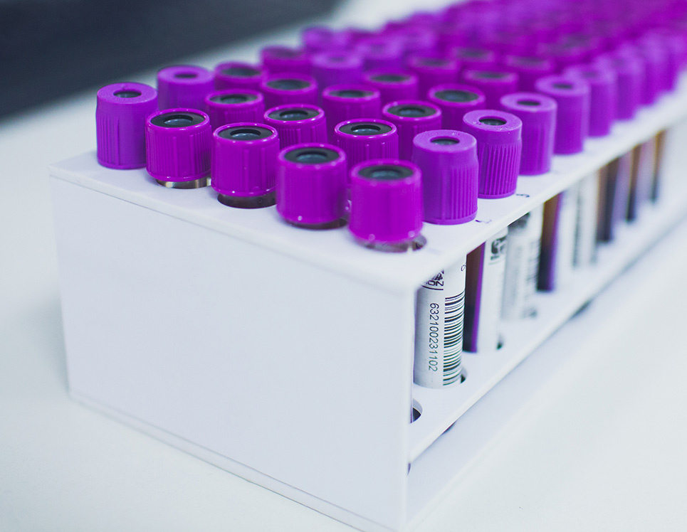
To begin with, the technology of blood sampling has now completely changed. In place of scarificators and glass capillaries with test tubes came vacuum containers. The blood collection systems now used are less traumatic, the process is completely unified, which significantly reduced the percentage of errors at this stage. Vacuum tubes containing preservatives and anticoagulants allow blood to be stored and transported from the collection point to the laboratory. It is thanks to the emergence of a new technology that it has become possible to pass tests as conveniently as possible - anytime, anywhere.
At first glance, it seems impossible to automate such a complex process as the identification of blood cells and their counting. But, as usual, all ingenious is simple. The basis of the automatic analysis of blood are the fundamental physical laws. The technology of automatic cell counting was patented back in 1953 by the Americans Joseph and Wallace Coulters. It is their name that stands in the name of the world brand of hematology equipment Beckman & Coulter.
Cell count
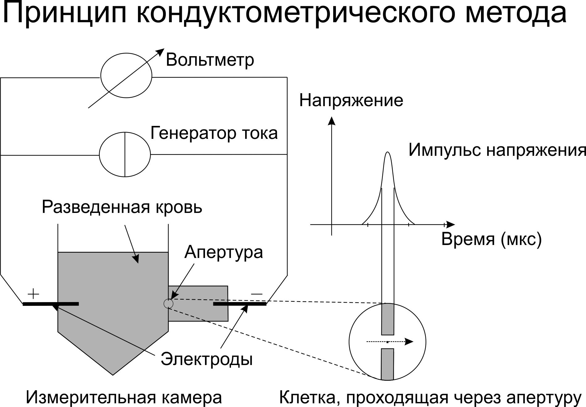
The aperture-impedance method (the Coulter method or the conductometric method) is based on counting and assessing the nature of the pulses that occur when a cell passes through an orifice of small diameter (aperture), on both sides of which two electrodes are located. With the passage of the cell through a channel filled with electrolyte, the resistance to electric current increases. Each passage of the cell is accompanied by the appearance of an electrical impulse. To find out what the concentration of cells is, it is necessary to pass a certain sample volume through the channel and count the number of pulses that have appeared. The only limitation is that the concentration of the sample must ensure that only one cell passes through the aperture at each time point.
Over the past 60 years, automatic hematological analysis technology has come a long way. Initially, these were simple cell counters that determine 8-10 parameters: the number of red blood cells (RBC), the number of leukocytes (WBC), hemoglobin (Hb), and several calculated ones. These were the first class analyzers.
The second class of analyzers has already determined up to 20 different blood parameters. They are significantly higher in terms of leukocyte differentiation and are able to secrete granulocyte populations (eosinophils + neutrophils + basophils), lymphocytes and the integral population of middle cells, which included monocytes, eosinophils, basophils and plasma cells. This differentiation of leukocytes was successfully used in the examination of healthy people.
The most technologically advanced and innovative analyzers today are third-class machines, determining up to hundreds of different parameters, conducting extensive cell differentiation, including maturity, analyzing their morphology and signaling to the laboratory doctor about the detection of pathology. Machines of the third class, as a rule, are also equipped with automatic systems for preparing smears (including their coloring) and displaying the image on the monitor screen. Such advanced hematology systems include the BeckmanCoulter equipment, in particular the UniCel DxH 800 cell analysis system .
Modern BeckmanCoulter devices use the method of multiparameter flow cytometry based on patented VCS (Volume-Conductivity-Scatter) technology. VCS technology involves estimating the cell volume, its electrical conductivity and light scattering.
The first parameter, the cell volume, is measured using the Coulter principle based on the evaluation of resistance when the cell passes the aperture at a constant current. The size and density of the cell nucleus, as well as its internal composition is determined by measuring its electrical conductivity in high-frequency alternating current. The scattering of laser light from different angles provides information about the structure of the cell surface, the granularity of the cytoplasm, and the morphology of the cell nucleus.
The data obtained on the three channels are combined and analyzed. As a result, cells are distributed in clusters, including separation according to the degree of maturity of erythrocytes and leukocytes (neutrophils). Based on the obtained measurements of these three dimensions, a number of hematological parameters are determined - up to 30 for diagnostic purposes, more than 20 for research purposes, and more than one hundred specific calculated parameters for highly specialized cytological studies. Data is visualized in 2D and 3D formats. A laboratory assistant working with a BackmanCoulter hematology analyzer sees the results of the analysis on a monitor in approximately the following form:
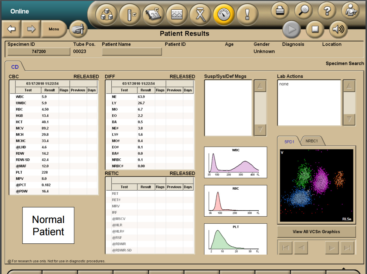
And then decides whether to verify them or not.
Is it worth saying that the information content and accuracy of modern automatic analysis are many times higher than manual ones? The performance of machines of this class is about hundreds of samples per hour when analyzing thousands of cells in a sample. Recall that during a smear microscopy, only 100 cells were analyzed by a doctor!
However, despite these impressive results, it is microscopy that still remains the “gold standard” of diagnostics. In particular, when the device detects a pathological cell morphology, the sample is analyzed under a microscope manually. When examining patients with hematological diseases, microscopy of a stained blood smear is done only manually by an experienced hematologist. In this way, manually, in addition to automatic cell counting, the evaluation of the leukocyte formula is performed in all pediatric blood tests on orders made using the laboratory online service LAB4U.RU.
Instead of a resume
Technologies of automated hematological analysis continue to actively develop. Essentially they have already replaced microscopy when performing routine prophylactic analyzes, leaving it for particularly significant situations. We are referring to pediatric tests, analyzes of people with confirmed diseases, especially hematological ones. However, in the foreseeable future, and in this area of laboratory diagnostics, doctors will receive devices capable of independently performing morphological analysis of cells using neural networks. By reducing the burden on doctors, at the same time, they will increase the requirements for their qualifications, since only atypical and pathological cell conditions will remain in the decision-making area.
The number of informative blood test parameters that have increased many times raises the requirements for professional qualifications and the clinician who needs to analyze combinations of the mass parameters for diagnostic purposes. To help the doctors of this front go expert systems, which, using data from the analyzer, provide recommendations for further examination of the patient and give a possible diagnosis. Such systems are already represented on the laboratory market. But this is a topic for a separate article.
Source: https://habr.com/ru/post/328508/
All Articles