Part 2. How many megabits / s can be passed through the optic nerve and what is the resolution of the retina? Some theory
Other publications in this series
Part 1. Unboxing VisuMax - femto laser for vision correction
Part 3. Meet - laser called Amaris. Moving and first awakening of VisuMax
Part 4.1 We return the sight. From points to the excimer laser
Part 4.2 We return the sight. From points to the excimer laser
The previous publication on laser vision correction technology was greeted with an interest, which I honestly did not even expect. That is why I decided to continue the article in the form of a whole cycle, within the framework of which we will examine in more detail the technologies underlying laser ophthalmosurgery. If you expected to see directly the lasers themselves in this article - I will disappoint you a little. For a long time I tried to circumvent the biological theme, but in the end I realized that I couldn’t talk about laser vision correction without revealing the basis of the structure and functioning of our vision.
')
I will try to consider human vision through the prism of IT. If someone is not too interested to read the part on the biological aspects of vision - do not worry. Just skip sections, starting with the optical system of the eye, and immediately proceed to the traditional competition from our girls. However, I would still recommend reading this material in order to better understand the following article, in which we will look at LASIK, Femto-LASIK, ReLEx SMILE and other methods of laser ophthalmic surgery.
There is a mood to figure out what exactly these strange white-labeled people say, looking thoughtfully at the results of your examination? Do you want to learn a little about the unique natural gift - vision? Then welcome to habracut. As usual there are a lot of illustrations and traffic (≈5 MB).
Table of contents:
Vision and IT
Optical system of the eye
Why do we see badly? Refractive disorders
Bonuses
Instead of intro
The eye is the most complex optical instrument created by nature. Man in many ways was just an imitator, creating his creations. Optics has come a long way as a science from Newton's experiments with a prism to the development of unique lasers, night vision devices and other interesting things. However, let's take a closer look at what inspired humankind to research in many ways and became the prototype of many modern things. The human eye.
Retina and optic nerve
Any device capable of recording light usually has one or another version of the photosensitive matrix. Its biological analogue is the reticular membrane of the eye.

Normalized plots of sensitivity of human cone cells of various types (K, C, D) and rod cells (P) to different parts of the spectrum. The wavelength axis on this graph is logarithmic.
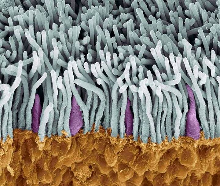
Electron micrograph of the retina. The rods are gray and the cones are purple. The illustration shows a clear predominance of monochromatic sticks over cones
For example, the retina of the human eye has about 7-8 million cones, which are responsible for color vision, and about 120 million rods (black and white vision). It would not be entirely correct to equate a cone / stick to a pixel, but, according to rough estimates, their number approximately corresponds to 250 megapixels for a panorama from both eyes. After excitation of the photosensitive cells of the retina, the signal must be transmitted to our biological CPU / GPU. This function is successfully performed by the optic nerve. The bottleneck against which the maximum frame rate transmitted by the organ of vision resists is the latency of the nerve synapses (the site of communication between neurons where the impulse is transmitted through the release and capture of chemical substances). According to various estimates, this is approximately 100-150 Hz, which is the limit of the speed of image transmission to the visual cortex of the brain. The number of nerve fibers in the optic nerve is approximately 1,200,000. If we assume that one fiber per cycle can transmit 1 bit of information, then the total throughput of the optic nerve is approximately 1.2 * 10 6 * 150 bits = 180 megabits / s. Solid stream. Our brain has to process a total stream of 360 megabits / s only from the visual analyzer. But there is still a rumor, smell, touch, temperature and pain receptors, a sense of balance, a proprioceptive feeling (feeling of one’s own body in space). All this load, not counting the heaps of other functions, fits into only 25 W of TDP.
Here, by the way, lies a number of issues that lie in the field of bioinformatics. For example, it is not entirely clear how ≈125 * 10 6 receptor cells can transmit information to ≈ 1.2 * 10 6 conducting neurons. That is, before transmission to the brain, some sort of filtering and preprocessing of visual information occurs. By the way, it follows from this that the retina is not able to transmit more than 1.2 megapixels for the “tact”. Another thing is that as a result of processing a series of such "snapshots" a much more distinct picture of what is happening is formed in the brain. Another interesting feature of human vision is the fact that we always see the past . The delay in conducting nerve impulses to treatment centers is, according to various estimates, about 150-180 ms. That is why, by the way, in professional sports is considered a premature start jerk athlete in the interval from 0 to 100 ms. It is believed that a person could not have time to react so quickly due to physiological limitations. It must be said that these values may vary within certain limits depending on stress, psycho-emotional state, the level of neurotransmitters, but on the whole the picture is quite stable.
Compare with the camera?

Photosensitivity can vary in the broadest range - from normal vision with illumination of 25,000 lux (bright midday) to the registration of individual photons in total darkness with maximum adaptation. The dynamic range of the eye also strikes against the background of traditional cameras - about 24 f-stops! For comparison, the maximum dynamic range among photographic materials has a black and white film - about 10 f-steps. The color film has a range of about 7, and the average matrix of a modern camera is from 4 to 6. Thus, the dynamic range of the eye is 2 (24-6) = 262 times the average camera.
It is worth noting that such an adaptation takes time. The habituation time is less when moving into a bright room - just a few seconds. When going into darkness, this time is extended to several minutes. This is due primarily to the need for the synthesis of destroyed rhodopsin , the visual protein, which is directly responsible for the occurrence of visual arousal. The luminous flux is regulated in the same way as in cameras - a diaphragm. This function is performed by the iris, the hole in which is called the pupil. Knowing that the focal length of the eye is about 22 mm, one can calculate the f-number for the pupil: for a maximally dilated pupil of 8 mm - 22/8 = 2.75, for a maximally narrow one - 22 / 1.1 = 20.
In fact, our eyes are a wide-angle lens with inherent distortions on the periphery. However, when the three-dimensional image is reconstructed in the brain, this is compensated. Also, we do not notice the blind spot , despite its quite tangible angular dimensions (about 1.3º).
Optical system of the eye
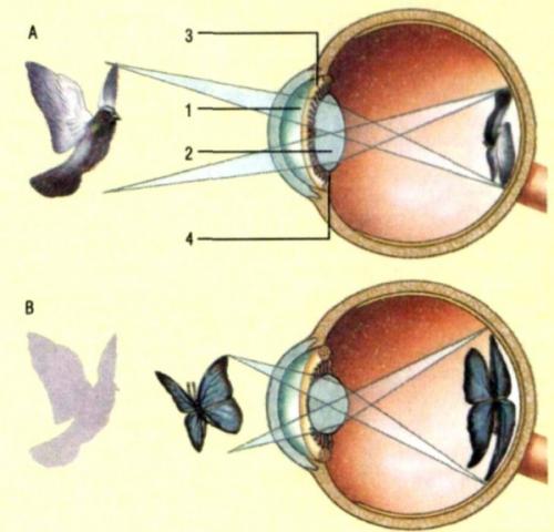
For correct focusing of rays, not only the size and shape of the elements, but also their coefficient of light refraction is important to us. It should be understood that each light guide element has its own, well-defined factors. By the way, this explains why we, unlike fish, see so poorly under water - our eyes have evolved as an organ to ensure clear vision on land. All this happens because the optical power of the lens depends on the refractive index of the medium.
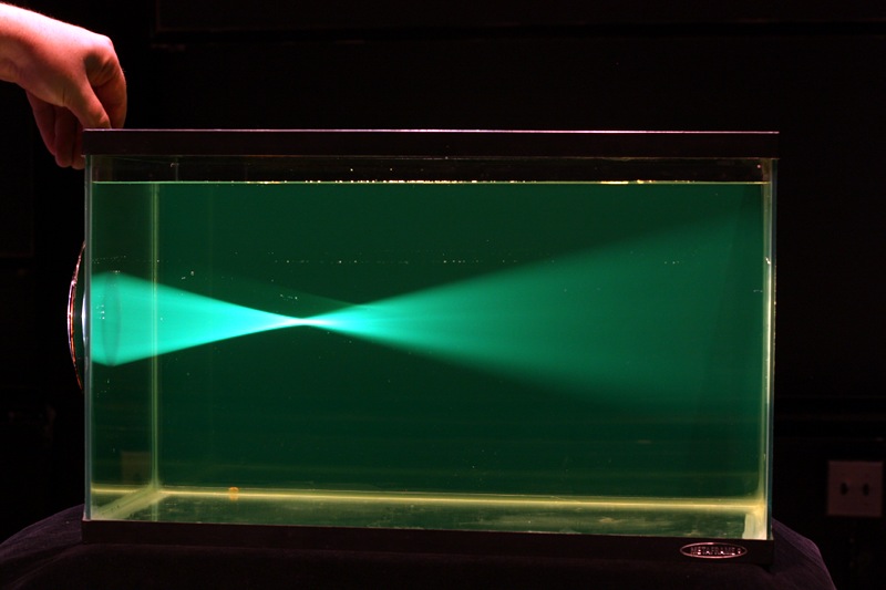

Glass lens significantly loses the ability to focus light when transferred to a medium with a different IOR
The eye is a finely balanced optical system. The slightest changes in the ratio of its elements lead to a violation of the final image on the retina. Imagine for a moment that you took and put in manual mode an arbitrary focus on the camera lens. Agree that a good and clear picture in this way you will not get. Consider in more detail which path overcomes the light on the way to the retina.
Optical system of the eye. The main dimensions and refractive indices of the media are indicated.
Cornea
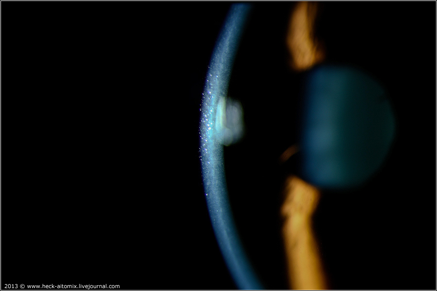
Cornea with slit ophthalmoscopy
Cornea with high magnification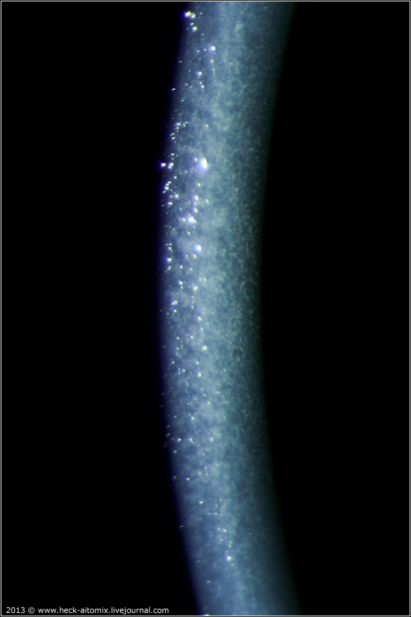
The first element that meets the onslaught of a not-too-friendly environment is the cornea. The cornea belongs to the outer shell of the eye and performs a protective and light refractive function. It is this shell of the eye that is confronted with the constant negative impact of dust, speckles, sunlight and many other factors. The epithelium of the cornea is constantly updated, ensuring its integrity and protective properties. Interestingly, the cornea does not have blood vessels in its optical part - this would violate the transparency. Food and gas exchange of these areas is carried out by diffusion due to tear fluid from the outside and aqueous humor of the anterior chamber from the inside.
Front and rear cameras
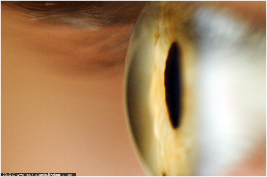
The photo shows the anterior chamber of the eye, located immediately below the cornea and filled with a special liquid — aqueous humor. The rear camera is located behind the iris and pupil.
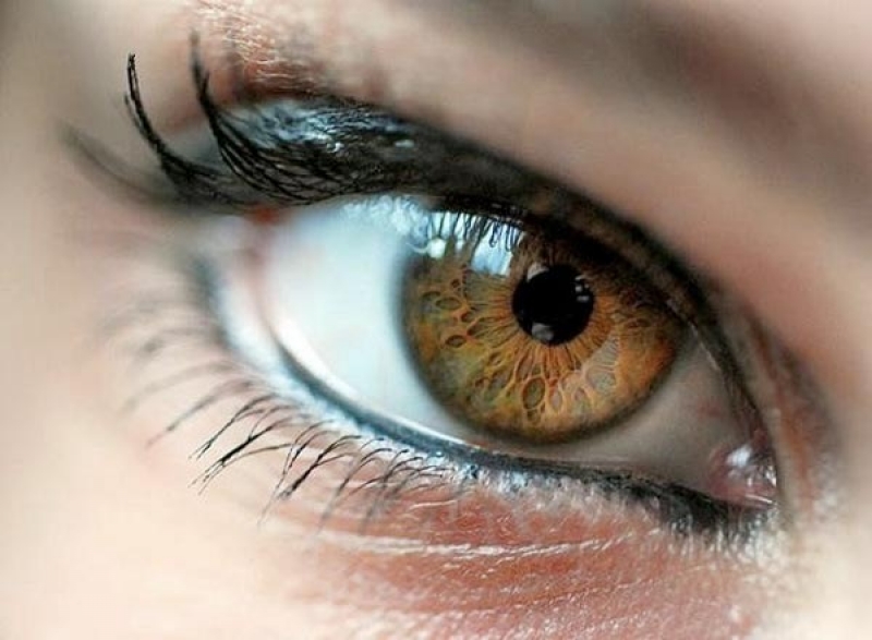
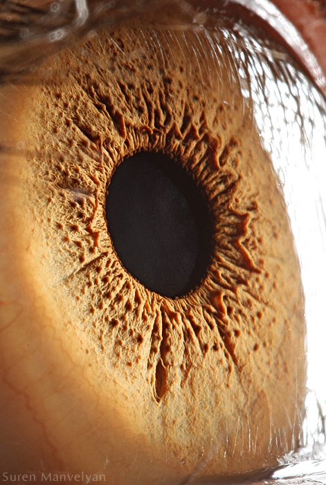
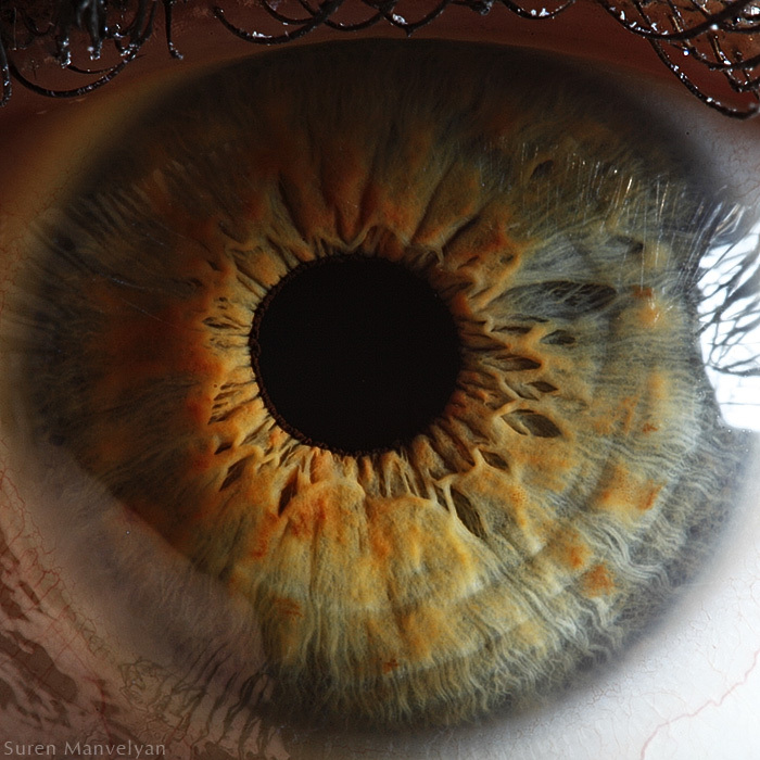
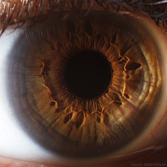
Iris and pupil
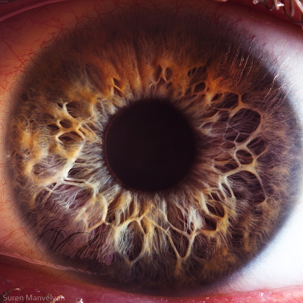
Each person is the owner of the iris pattern unique in its beauty and palette. For an IT specialist, iris is first and foremost an important element for biometric identification. For the doctor, this is a very important element that, like the diaphragm, regulates the flow of incoming light. The hole in the iris is called the pupil . Few guessed that the iris has a fairly pronounced three-dimensional relief, which can be shown in the pictures with the correctly set light. Polarizing filters are often used to suppress corneal flare.
By the way, many have never thought that the dependence of the depth of field image and the diameter of the diaphragm extends to the pupil. In the dark, the depth of field decreases sharply as the pupil increases in aperture.
More beautiful photos of the iris.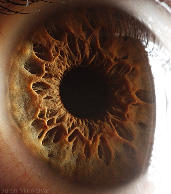
Lens
The lens is a unique biological lens that has a number of extremely important properties. One of the most important is the ability to change its curvature under the influence of the ciliary muscle. This process is called accommodation and allows you to focus on both remote and very close objects. The accommodative capabilities of the optical system of a young man’s eyes are ~ 14 diopters, gradually diminish with age and are almost lost by 60–65 years. By the way, it is thanks to the lens that the optical system of the eye is so compact compared to, say, SLR cameras.
The reflection of the front surface of the lens with slit microscopy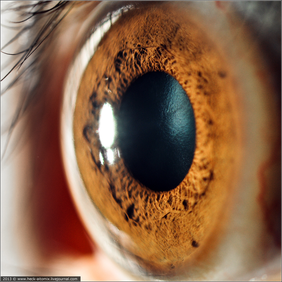
Vitreous humor

Contrary to popular belief that the eye is filled with fluid that can leak out at the slightest puncture, the main volume of the eye is occupied by the vitreous body. This substance is more like a viscous gel, whose mechanical properties are determined mainly by hyaluronic acid. The main function of the vitreous body is to maintain the stable shape of the eye, giving it the necessary elasticity. Also, the vitreous body passes through itself and refracts light.
Refractive disorders
All of the above elements refer to the refractive system of the eye. That is why, any violations associated with them are called refractive . This type of pathology is interesting because we have the ability to restore the correct course of the rays by acting not on the element that caused the disease. For example, the use of glasses as an additional lens, corrects myopia, the cause of which was an increase in the central optical axis of the eye. Consider further the main problems associated with refraction.
Hobragitel ansaril3 proposed to add to the article the physical justification of such violations. Unfortunately, my medical education does not allow me to fully understand the meaning of such things, but I will leave a link for those who are interested.
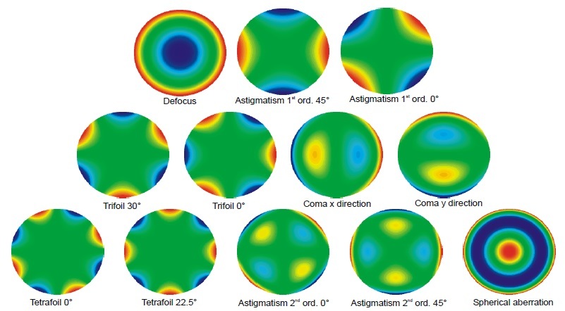
Zernike polynomials and wave aberrations :
Myopia (myopia)
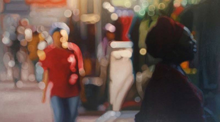
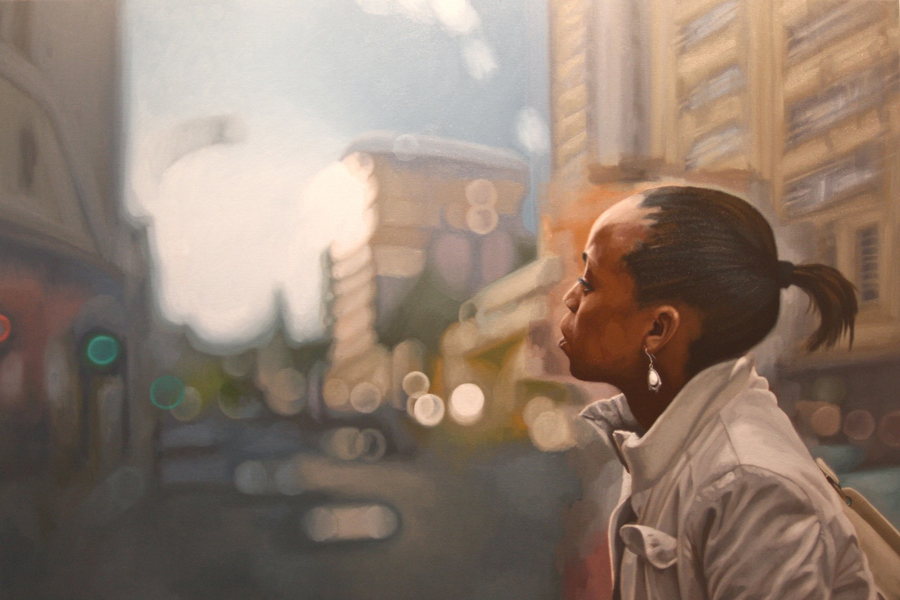
The scourge of many experts in the field of IT. Before talking about the causes of this disease, I want to briefly turn to art. Philip Barlow is a talented South African artist who was able to reflect the world through the eyes of a myopic man in his works.

Some more works by this author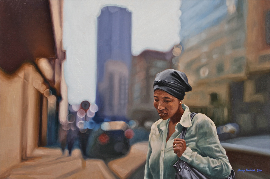

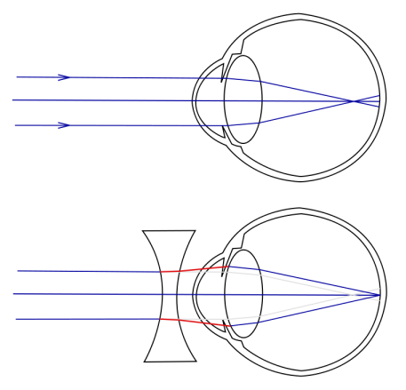
The cause of myopia is an increase in the size of the eyeball along its optical axis:
This disease is most often manifested in adolescence, during a period of rapid growth of the body. There are observations that link the excessive stretching of the eye with genetic disorders in the synthesis of collagen. Collagen is a structural protein that is important in the formation of connective tissue. With its excessive elasticity, a disproportionate growth of the eyeball occurs. At the growth stage, sclerotherapy and collagenoplasty can be used to stop further growth of myopia. The essence of these methods is to increase the strength of the outer shell of the eye - the sclera - due to the implantation of a special material. The treatment of this pathology consists in non-operative methods (glasses, contact lenses) and surgical methods (various types of laser vision correction).

We have the possibility of correcting a similar problem with the Femto-LASIK technology, in the coming weeks we will also be able to work with the ReLEx SMILE technology. We use the latest generation of surgical lasers:
For Femto-LASIK, this is Amaris 750S from Schwind (excimer laser for correction itself) and VisuMax from Zeiss (femto laser for cutting out flap)
For ReLEx SMILE, this is only VisuMax (technology implies performing all manipulations only on it, which is discussed in more detail in the next article)
Accommodation spasm
It is necessary to distinguish between true myopia and the so-called spasm of accommodation , which is also false myopia. I think that by virtue of the profession, many of us spend an enormous amount of time continuously looking at the monitor. As we said earlier, the ciliary muscle is responsible for the accommodation, which deforms the lens in the necessary way. With constant visual tension, when the focus of view is near for a long time, this muscle experiences a spasm and cannot relax. As a result, the eye loses the ability to normally focus on objects away, but this is due to temporary phenomena in terms of accommodation, and not to a change in the shape of the eye. In such a situation, special drugs are prescribed that cause temporary paralysis of the ciliary muscle, helping it to relax (tropicamide, atropine, and others). Visual gymnastics and observance of occupational health (illumination of the workplace, breaks, etc.) will not be out of place
Astigmatism

This pathology is often combined with others. Its cause is the asymmetry of the curvature of the cornea or lens. The result is a different birefringence with respect to different axes. As a result, a person can clearly see, for example, horizontal lines, while vertical lines will be blurred.
A bit more information about astigmatism.
Type of test world through the eyes of a person suffering from astigmatism

Test world in its original form.
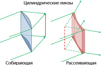
Treatment - the use of special cylindrical lenses in glasses or laser vision correction, during which this pathology will be corrected.
Type of test world through the eyes of a person suffering from astigmatism
Test world in its original form.
Treatment - the use of special cylindrical lenses in glasses or laser vision correction, during which this pathology will be corrected.
Hyperopia (hyperopia)

The reverse condition of myopia. The optical axis of the eye is shorter than it should be, with the result that the image is focused behind the retina.
This disease is often confused with presbyopia (age hyperopia)
Treatment with ophthalmic lasers is almost the same as with myopia.
Presbyopia (age hyperopia)
The peculiarity of this refractive violation is that a person loses the ability to accommodate with age. The lens becomes more rigid, the ciliary muscle is harder to deform. As a result, what is sometimes jokingly called “short hand syndrome” develops (This is not my bad vision, but my hands are short!). The lens is fixed in the “focus on infinity” position and loses the ability to accommodate close objects. As a treatment, a person replaces a natural mechanism for wearing glasses when it is necessary to consider something close. It is clear that we are not talking about accurate dynamic calibration of optical power. There are complex variants of progressive lenses, a number of other methods, but in any case this is not a complete replacement of the natural mechanism.
Practice of laser ophthalmosurgery
In order to reveal in more detail the possibilities of surgical lasers for vision correction, I asked Alexander Alexandrovich Boyko to write a few words about one of our ophthalmosurgeons, cms. I am afraid that he will not have time to answer on his behalf on Habré due to his constant workload, but, at my request, he took a few minutes to share his impressions here:
I have always said that the doctor stops being a professional at the moment when he stops learning something new. It is not a secret for any of you that medicine does not stand still, and every year new methods appear that allow for a more complete and high-quality treatment. When I chose my profession as an ophthalmologist, it was even difficult for me to imagine the technologies I would have to work with in the future. I realized that the future had already come when I was trained in 2013 to work with our new ophthalmologic lasers.
This is a training on working with the Zeiss VisuMax femtosecond laser in Jena, Germany, and the SCHWIND Amaris 500E excimer laser in Ashakhenburg. It is with great pleasure that I remember these few weeks of continuous study, professional teachers who told us about all the nuances of working with this truly wonderful equipment.
The ergonomics of these lasers are really thought out so that the machine becomes a natural extension of the hands and thoughts of the surgeon, allowing you to quickly and accurately perform therapeutic manipulations. The Amaris excimer laser allows for vision correction in a wider range and with even greater accuracy than its predecessors. Correction of myopia to -15 diopters, farsightedness to + 8D, as well as difficult cases of astigmatism to ± 7 diopters of the cylindrical component. And the VisuMax femto laser makes it possible to cut out a flap with a complex three-dimensional structure, which is calculated individually for each patient. This gave us the opportunity to expand the indications for surgery in people with a fairly thin cornea.
Together with our colleague, Ivan, we will try to prepare in the next article the most complete and detailed overview of all those methods and techniques by which we have a wonderful opportunity to return people a clear view of the world.
The article used materials:
- www.cambridgeincolour.com
- heck-aitomix.livejournal.com/67763.html with amazing images of slit ophthalmoscopy
- Amazing photos of the iris from Suren Manvelyan
- Textbooks on normal human physiology, ophthalmology.
Special thanks to the people who helped in editing the article and made their valuable comments:
- colleague Sunchea , who helped bring the material into readable form and gave a lot of valuable advice
- vvzvlad
- ansaril3
- DIHALT
- ,

Tanya-designer is working on the task))
Our female half of the team took into account the wishes regarding the complexity of the tasks and came up with something new)) This time more people joined the process, especially when it came to prizes. So, as a prize for the first 10 people who decide this quest certificate for a full tour of the clinic, with a visit to the server room, direct inspection of laser systems (during off-hours, of course) and a full range of vision diagnostics for the winner . I hope you will like it. I understand that many people live in other cities and will not be able to come, but you will be able to give someone a certificate (just let us know) or visit us if you are suddenly in our area (Krasnodar, Essentuki, Perm).

Small survey
There was an idea to hold a small webinar in which our technical director and one of the laser surgeons will participate. On it, everyone will be able to ask questions of interest that remain undisclosed in a series of articles. Accordingly, I want to learn from the habrasoobschestva opinion on this matter. It would also clarify which platform (free) is better and more convenient for most to use for a webinar.
UPD. Announcement of winners
I apologize that we delayed the winners, but not all the people who sent the letter gave us their nicknames in habrahabr. Publishing their e-mail would be wrong. As soon as we get them all - we will post the list. As a general assessment of the activity of the community, I can say that the first 10 answers were already an hour after publication. The counter shows approximately 300 clicks on the final quest link. About 40% of those who decided the quest were also able to find Easter eggs.
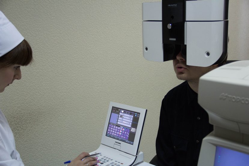
Coincidentally, one of the first guesses was vvzvladwho was not far from us and was able to get to our Krasnodar clinic. We honestly completed our part, combining the story of the diagnostic equipment with the conduct of the survey itself. And, of course, they could not help but touch the Femto-laser VisuMax, which I described in the previous article.

Source: https://habr.com/ru/post/207990/
All Articles