View from the inside: the world around us - 4
In the last article, among other things, it was mentioned that it was time to complete the epic with writing articles about the microcosm that surrounds us everywhere, but it was not there ...
')
One of my colleagues in electron microscopy, Andrei Burmistrov (@Venelt), who works on a serious Tescan microscope, periodically publishes on Magnomet.ru a great frame of microscopic objects, both living and non-living. And I wanted to share this wonderful world with respected users, let's go ?!
Inanimate matter
Let's start with the inanimate nature.
Carbon. New reading
In one of the articles I have already published carbon-fiber photographs or colloquially “carbon”, but I think it will not be superfluous to repeat, especially the photographs presented below were made “correctly”, with appropriate sample preparation.
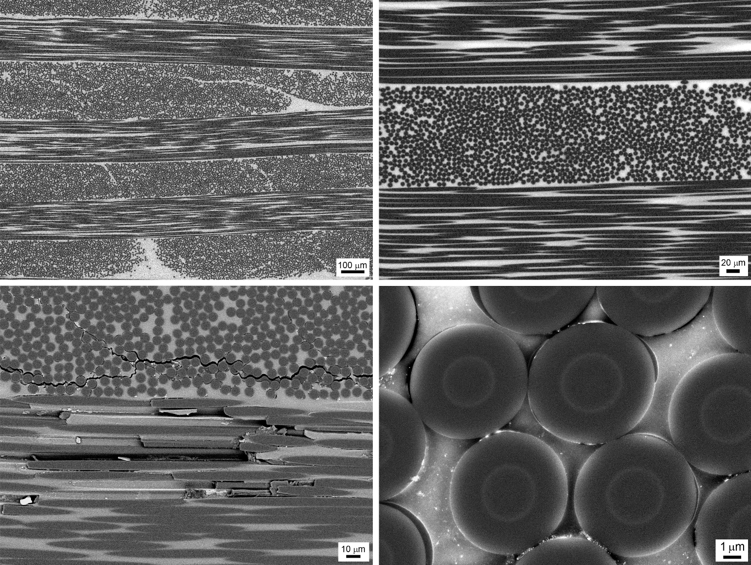
"Correct" micrographs of carbon fiber
These photographs are noteworthy in that they were obtained using a BSE ( back-scattered electrons ) detector, which allows you to see, for example, that the fiber is heterogeneous inside and consists of a core and a shell, which is apparently due to the conditions of fiber annealing.
Lighter sparks
I don’t think that anyone ever thought that each lighter of a lighter produces a huge amount of the smallest micro and even sometimes nanoparticles that fly in all directions. Often, they acquire completely bizarre forms depending on the conditions of formation, such as these:
But more, in my opinion, the particles are interesting, which during their short but bright life managed to crystallize, forming a unique pattern, according to which, in principle, one can suggest their phase composition:
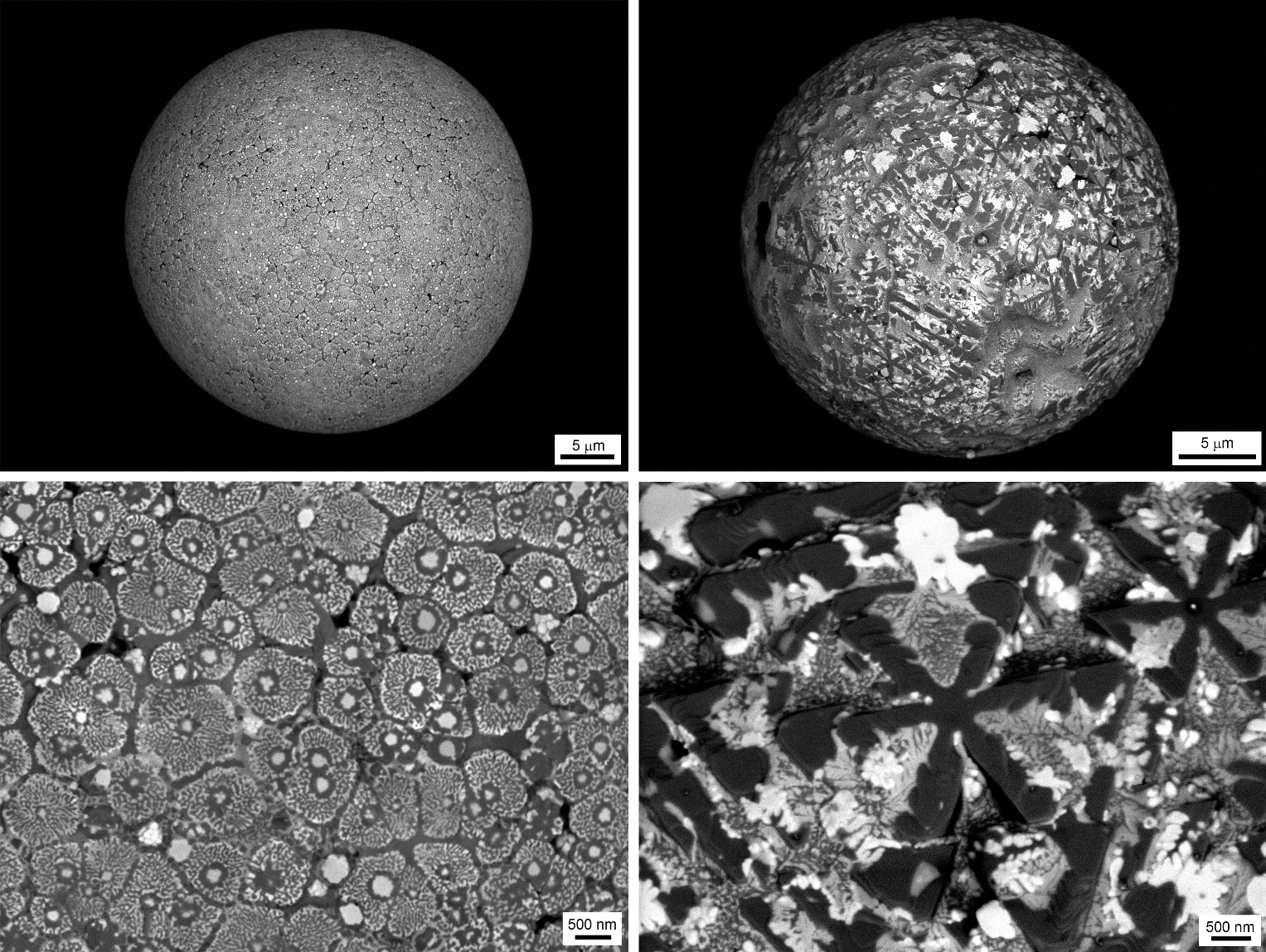
Images taken with a BSE detector giving information on the chemical contrast of the material (darker areas correspond to lighter elements)
But that's not all, due to the development of computing power of computers and processing electronics inside modern microscopes, it is now possible to get 3D (stereo pair) images of objects in one click. The electronics itself simply deflects the electron beam with which the surface is scanned at a given angle (usually 15-20 degrees), and then automatically combines two images:
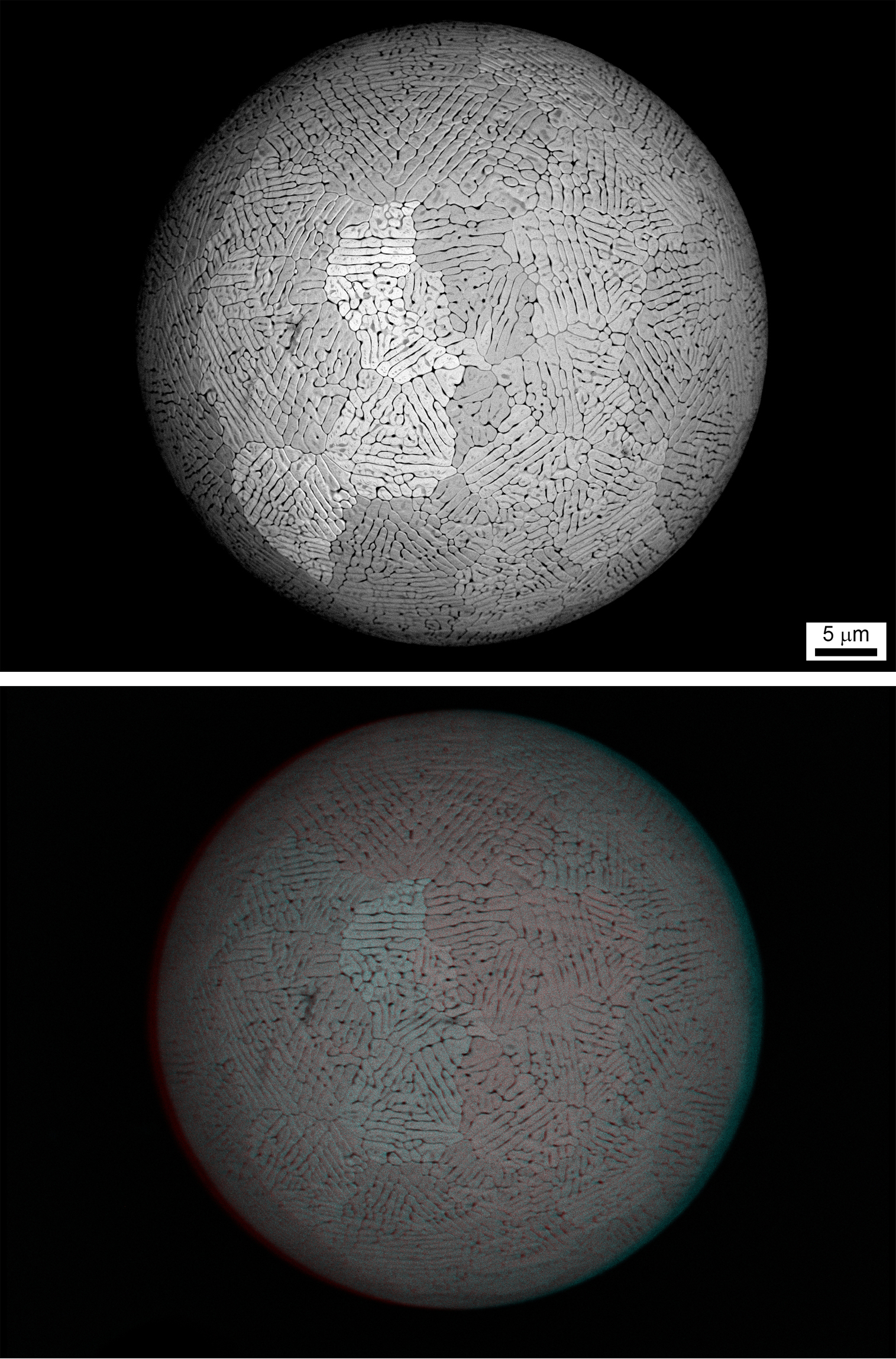
An example of using 3D in modern electron microscopy (requires glasses with a red and blue filter)
Toner
On Habrahabr, photographs of toner taken by scanning electron microscopy (for example, a post from Kyocera ) sometimes slipped. I hasten to add these wonderful pictures:
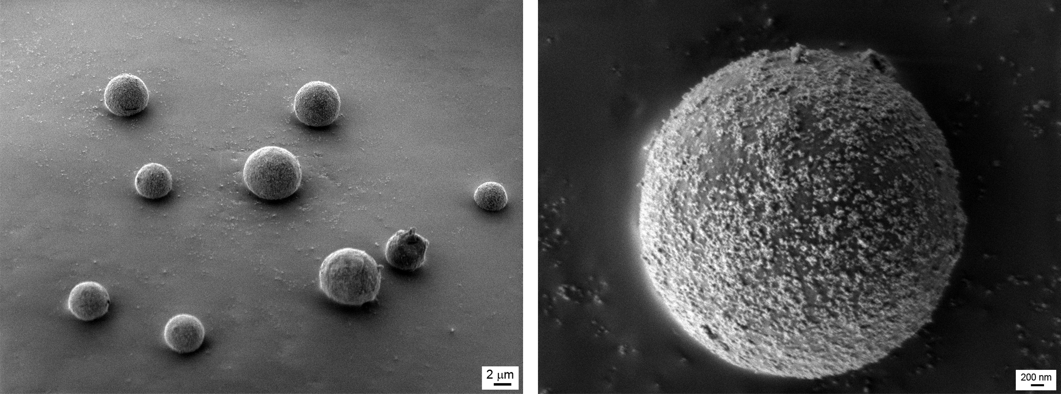
Spherical toner particles of printers usually do not exceed a few microns, so they are so difficult to remove if you get dirty.
Track membranes
Track membrane - a rather specific and outlandish thing. If you don’t go deep into details, you can get it by exposing (or exposing) a thin film of only a few tens of microns to ionizing radiation, for example, alpha particles (or accelerated inert gas ions). Penetrating through the film, alpha particles or ions because of their mass and speed fly through, leaving the track - a kind of well in diameter from a few nanometers to several microns (depending on the process).
After processing, such a sieve can be used to filter water, blood dialysis, fine air purification for clean rooms, and so on, up to creating clothes (I would not be surprised if GoreTex uses the same technology).
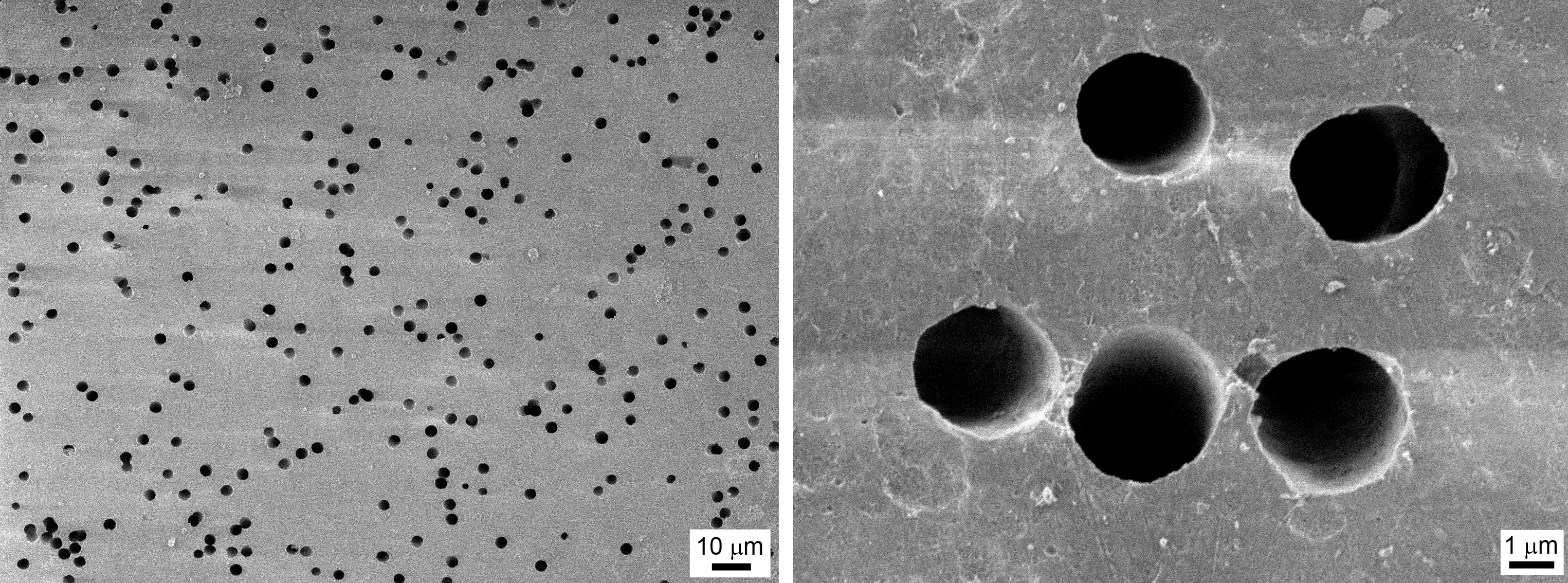
Lavsan track membrane (or, simply, PET )
Living matter
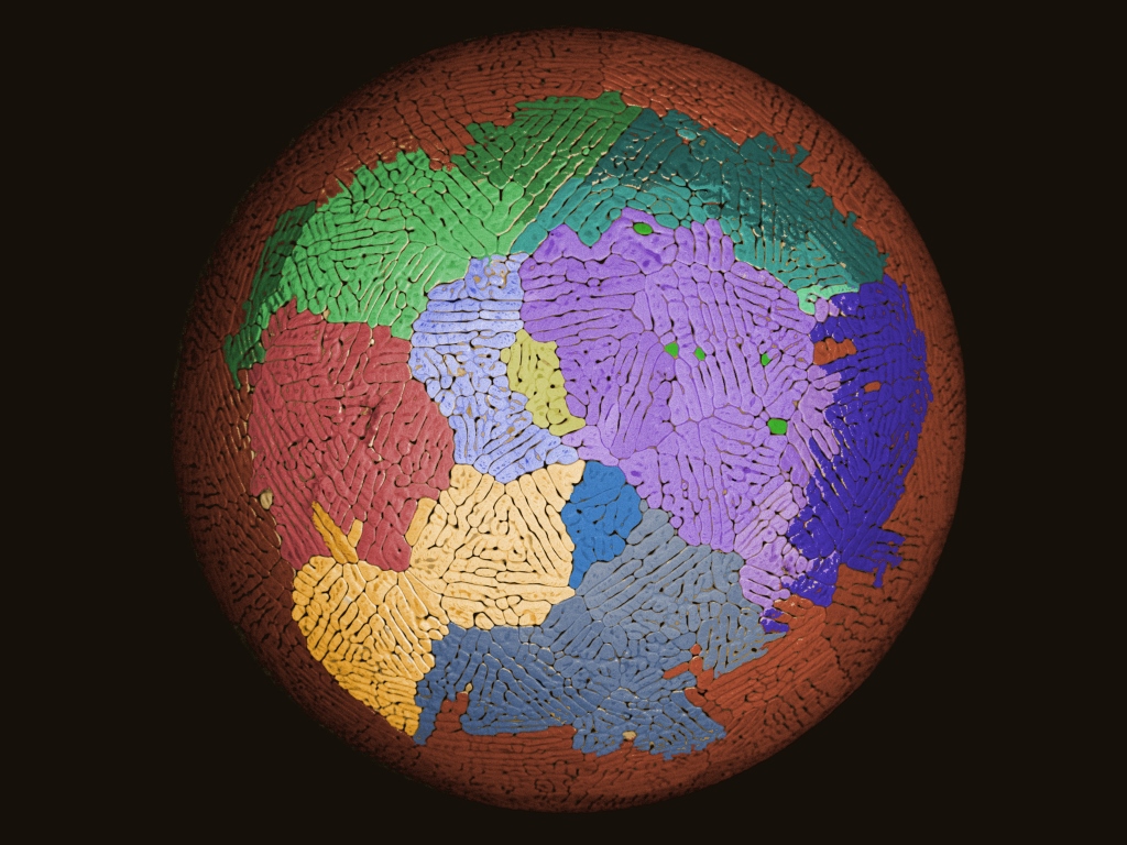
Butterfly wing
When it comes to optics, photonics and related fields of science, a butterfly and its unusual wings are immediately remembered. Exclusively due to the wave properties of light such structured wings can change their color depending on the angle of observation. Reasonable question: How does this wing work? And here is the answer:
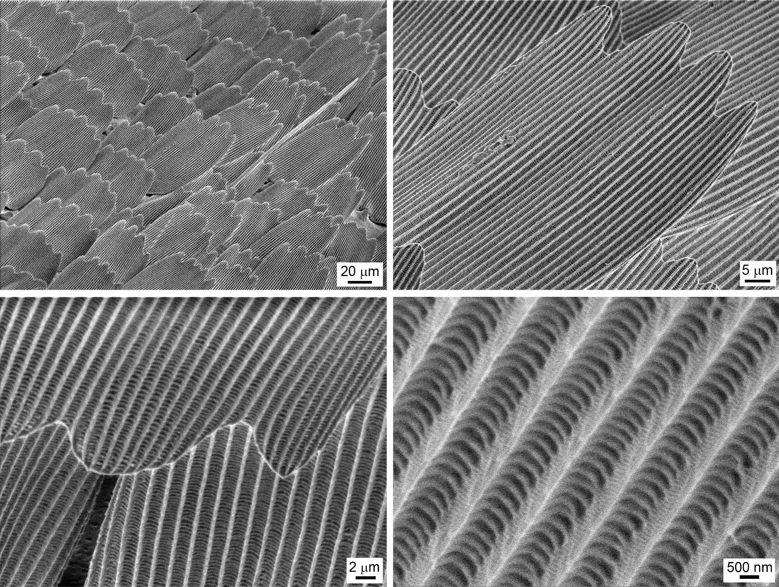
Butterfly wing under the electron microscope beam
As it is easy to see, the wing has several levels of structural organization. It consists of scales, which in turn consist of furrows. And each furrow is formed by a huge variety of small scales laid in a stack, the size of which exactly coincides with the wavelengths of the visible range, so we have a diffraction grating that sets the color to the wing of the butterfly. Consequently, depending on the angle of observation, we see one or another color.
Web
No one has seen this for sure, and I confess honestly, when I first looked at these photos, I simply could not understand what was in front of me. But it turned out that this is only a cobweb. And, perhaps, I would venture to suggest that these balls (with some sticky secretion of the glands, apparently) on it are places for attaching transverse filaments and / or a trap for flies and other insects. The thickness of the rope for the web about 1 micron!
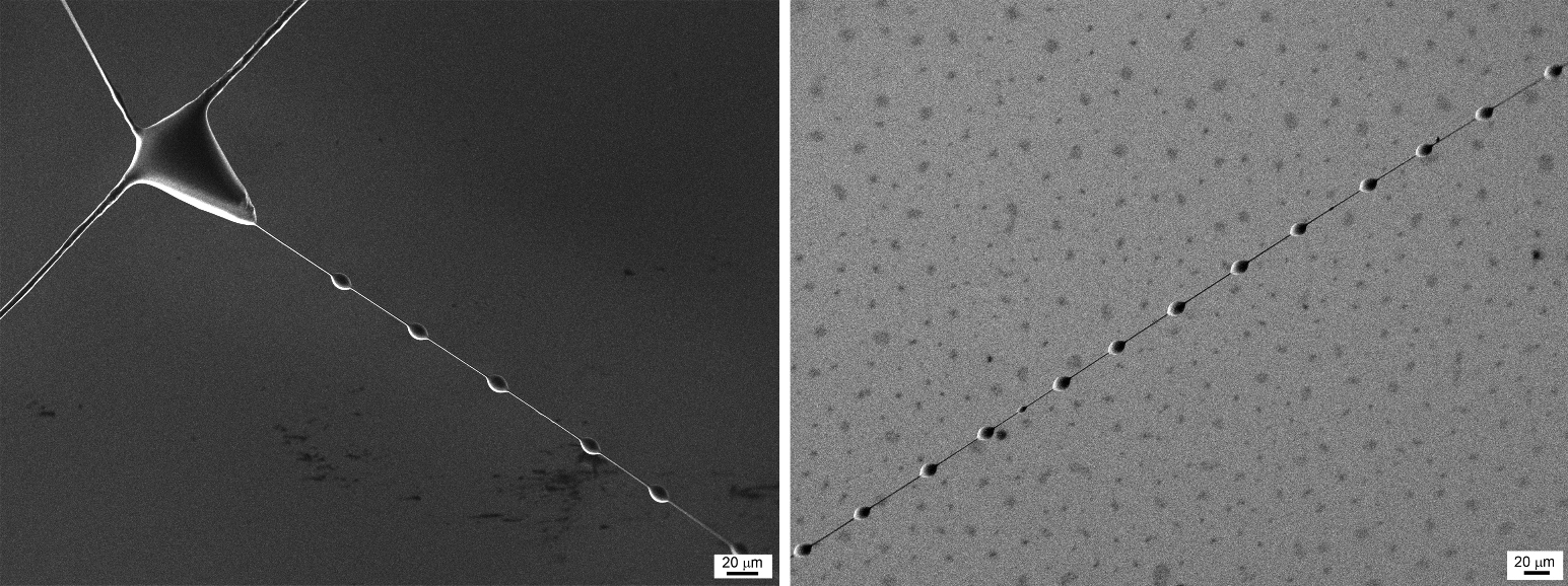
Ideally stretched strings of spider webs
Insect
By the way, about insects. It seems that in one of the early posts I was asked to show some insects at the point of an electron microscope. You know, in Russian fairy tales it usually sounds like this: “How long, how short is it ...”
In general, meet our guest - Muhu-Tsokotuha. We need to clarify with Andrew, but perhaps she was just the victim of the spider-arranged networks ...
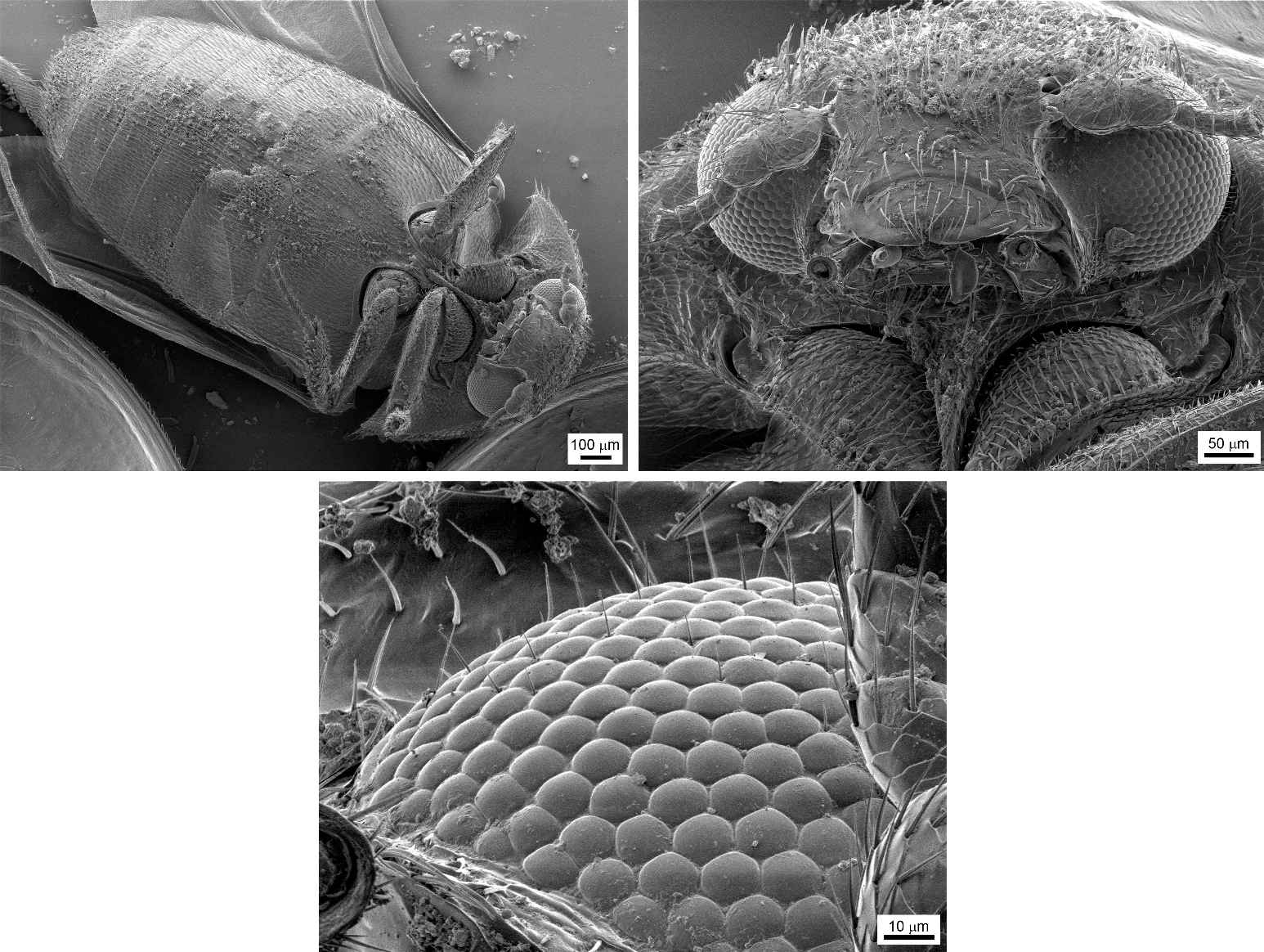
A fly under a microscope and its faceted eyes
Usually, since childhood, we are vaccinated with hostility to various insects, including flies, they say, if the fly has sat on some object, then before use it must be washed again. Now you, dear readers, have a clear confirmation of this - a lot of particles of dirt and, possibly, bacteria on the insect's legs:
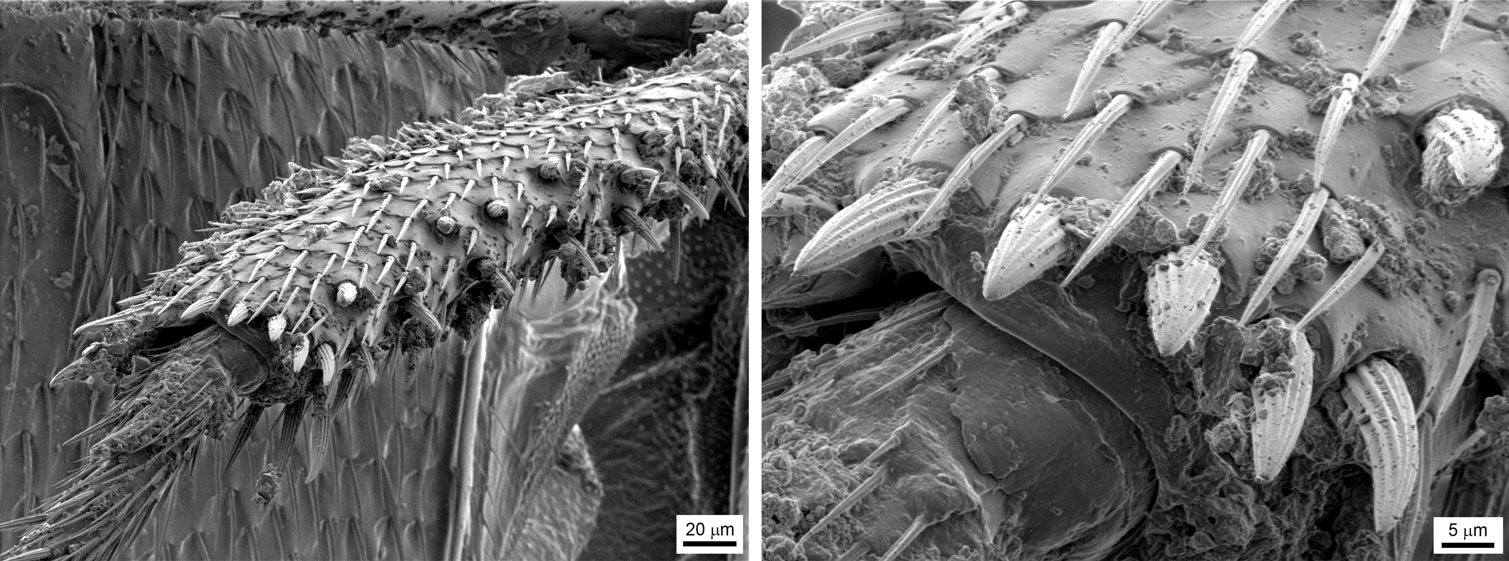
Increased fly's foot and dirt on it
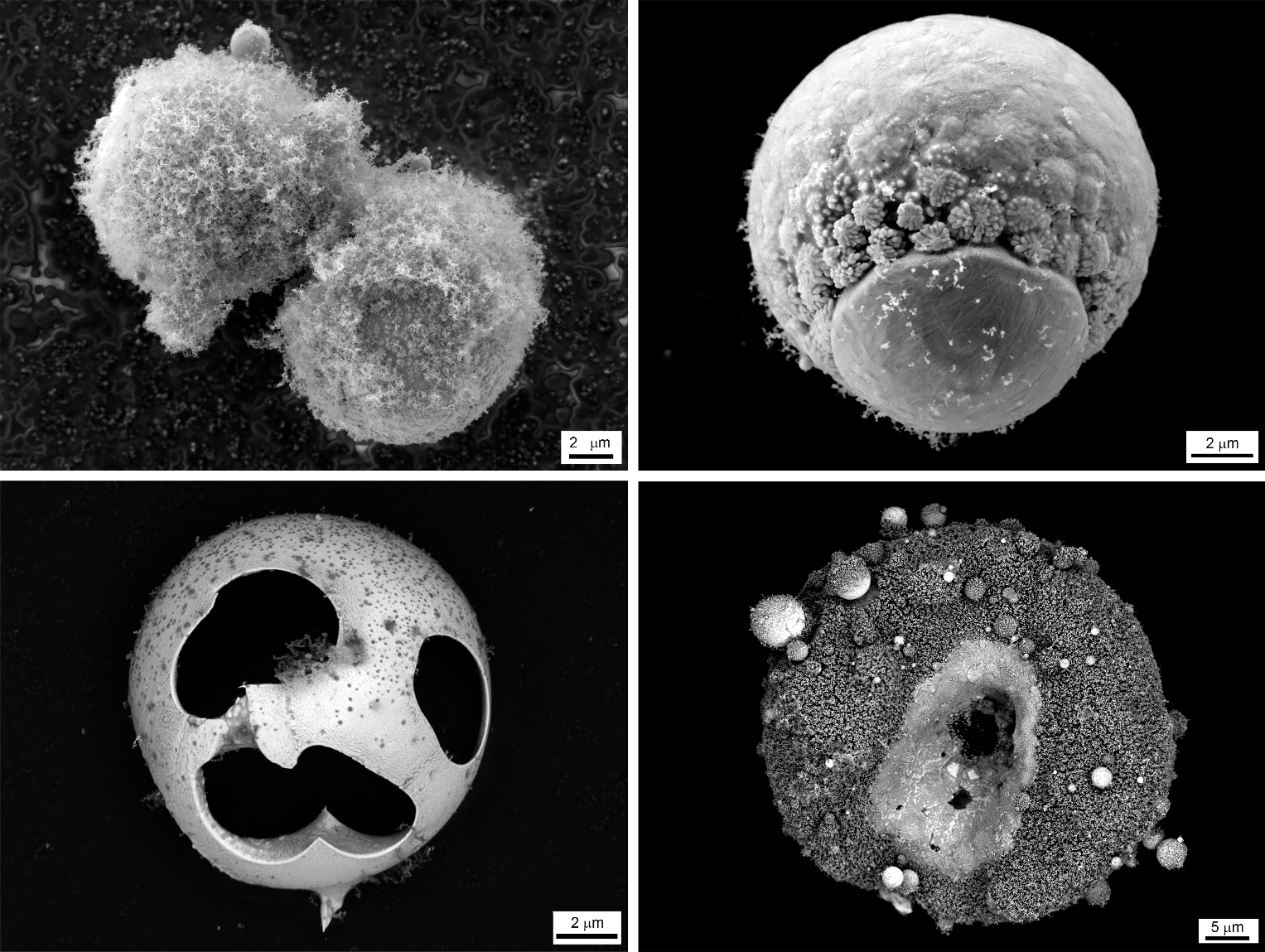
Radiolarians
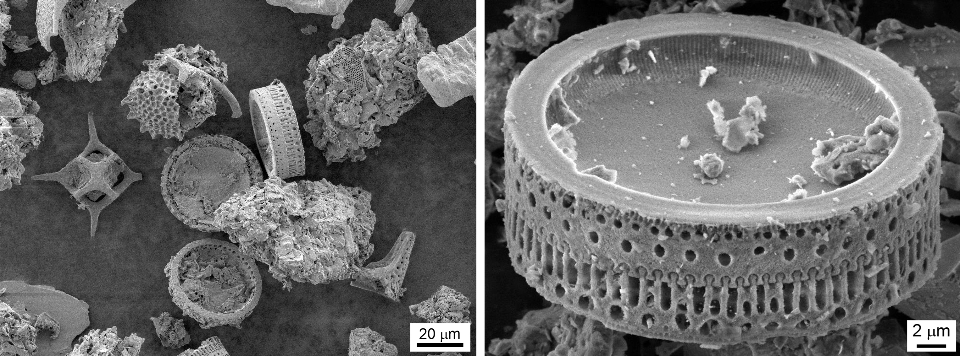
And finally, today's release completes another unusual object that surrounds us everywhere in the warm waters of the oceans - single-celled plankton or radiolaria . In principle, unicellular and unicellular are nothing special if it were not for their skeleton, which, by the way, after the death of these, forms bottom sediments ...
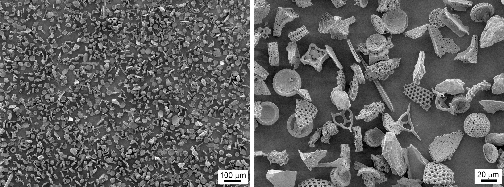
The designer of "Radiolaria" - collect what you can!
Sometimes in samples there are practically unbroken copies of the most bizarre forms:

Well preserved during transportation from the warm seas to Moscow
And a bit of intrigue in the style of REN-TV: it seems to me that the ancient Romans had an electron microscope and spied on the idea of the Colosseum in nature, that is, they were the founders of the science of biomimetics (a joke, of course, but involuntarily you start to think):
The Colosseum is now everywhere in the world ocean!
Finally - a journey from a millimeter to a micrometer - from a crustacean to diatom and further to bacteria:

PS: You can also say thanks to the author of photos Venelt .
First , the full list of published articles "A view from the inside" on Habré:
Opening the Nvidia 8600M GT chip , a more detailed article is given here: Modern chips - a view from the inside
An inside look: CD and HDD
An inside look: LED bulbs
An inside look: the LED industry in Russia
An inside look: Flash and RAM
An inside view: the world around us
Inside View: LCD and E-Ink Displays
An inside look: matrix digital cameras
An inside look: Plastic Logic
An inside look: RFID and other tags
An inside look: postgraduate study at EPFL. Part 1
An inside look: postgraduate study at EPFL. Part 2
An inside view: the world around us - 2
An inside view: the world around us - 3
View from the inside: the world around us - 4
An inside look: 13 LED lamps and a bottle of rum. Part 1
An inside look: 13 LED lamps and a bottle of rum. Part 2
An inside look: 13 LED lamps and a bottle of rum. Part 3
An inside look: IKEA LED strikes back
An inside view: are Filament lamps good for you?
and 3DNews:
Microview: a comparison of modern smartphones displays
Secondly , in addition to the blog on HabraHabr , articles and videos can be read and viewed on Nanometer.ru , YouTube , and Dirty .
Sometimes briefly, and sometimes not so much about the news of science and technology, you can read on my Telegram channel - welcome;)
Source: https://habr.com/ru/post/206296/
All Articles