Nokia: Now a fluorescent detector of nanoparticles and viruses

They won’t think up anything for owners of the most popular in the USA gadget from an apple company: optics for photographing, solar panels on the back panel for environmentalists, portable microprojectors and so on and so forth. For Android, the gadgets are somewhat smaller, but for Nokia they are almost completely absent. But scientists from the University of California (University of California) decided to fill this gap, keep up with the world of high technology and wondered: what will happen if you combine a serious device (a fluorescent microscope) with a smartphone ?!
In a published paper in the ACSNano magazine (quite a solid magazine, it is worth noting) a team of engineers, chemists and physicians presented a “field” platform for fluorescence microscopy for ... parapes ... Nokia PureView 808, which can be used to detect and visualize without leaving the ticket office , nanoparticles and viruses.
')
The basis of the device is the camera of the smartphone itself, with, as you know, good optics, to which is light and compact - if you can call it at all, 186 grams, after all - an opto-mechanical system. This module consists of several important components: a compact high-power LED (laser diode) operating at a wavelength of 450 nm to excite fluorescence, a thin-film interference filter (longpass filter) to reflect diffuse exciting radiation, several external lenses that magnify the image 2 times, and a movable frame for focusing.
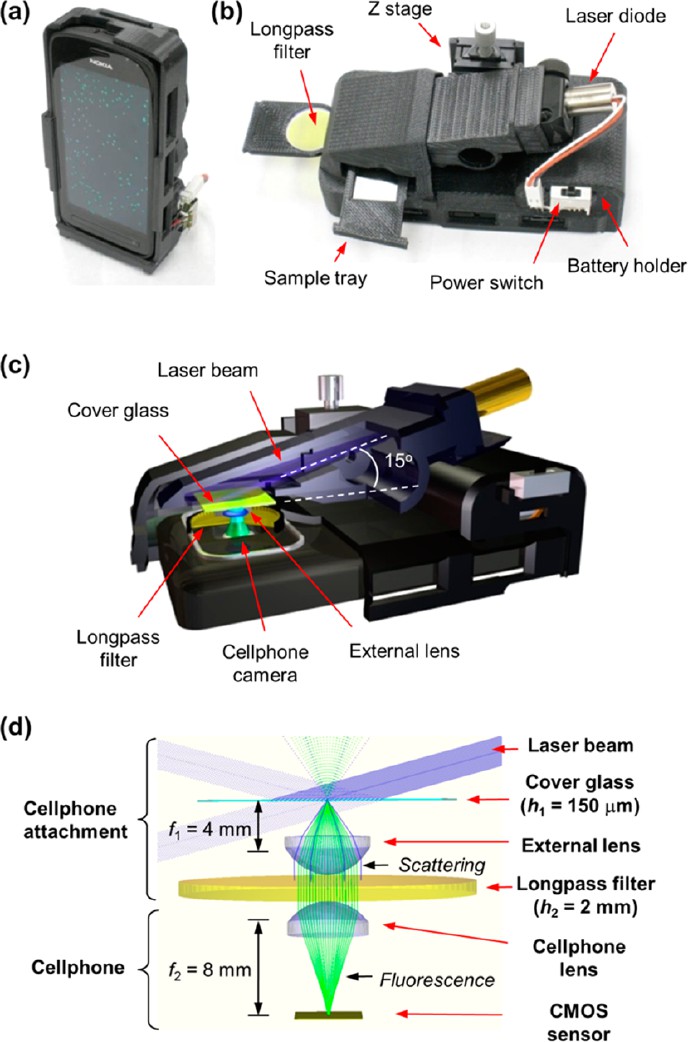
a, b) Photos of a smartphone with an attachment for optical fluorescence microscopy. c, d) Diagrams explaining the principle of operation of the device
The authors tested the device created by them on 100 nm of fluorescent nanoparticles, as well as on the most common virus (human cytomegalovirus ). Both nanoparticles and viruses were pre-immobilized on the surface of the specimen glass and also “tinted” with a special fluorescent label (in the case of viruses, these could be fluorescent antibodies ). Of course, the images obtained using the device were then compared with the original images of the same objects obtained using scanning electron microscopy (SEM) in order to confirm the correctness and correctness of detection. And also a number of tests were conducted to determine the resolution of the device.
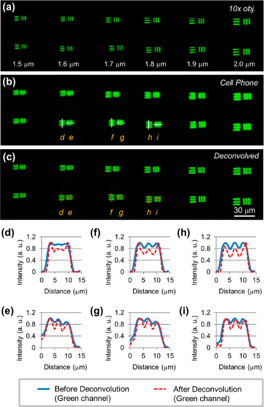
Prefix resolution test: a) image taken with a conventional fluorescence microscope, b, c) images of the same surface taken with Nokia and a profile along specified lines (di)
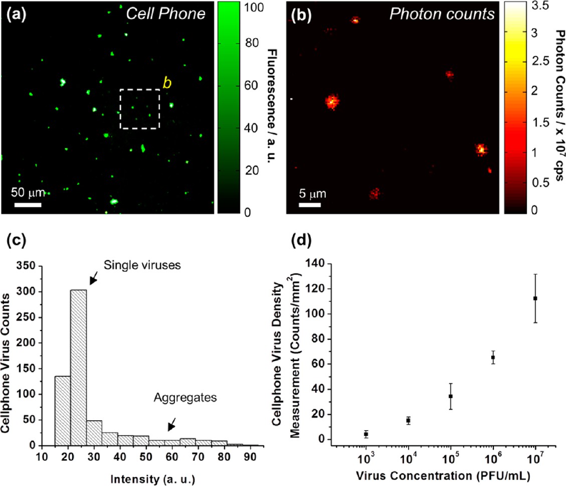
Detection of the virus and the dependence of the received signal on the concentration
The inventors themselves suggest using this device to visualize submicron objects (100 nm - 1 micron). This may include both bacteria and viruses, and after appropriate certification, it could be used “in the field” for measuring viral load and other biomedical tests and tests, especially in remote and resource-limited areas (for example, in Africa).
Sometimes briefly, and sometimes not so much about the news of science and technology, you can read on my Telegram channel - welcome;)
Source: https://habr.com/ru/post/203812/
All Articles