Homemade X-ray CT scanner
A month and a half ago, I talked about how Ben Krasnov (Ben Krasnow) collected a homemade X-ray scanner . Ben did not stop his experiments with X-rays and now represents a full-fledged computerized tomograph ( author's description ).
An example of the tomograph is shown below. Can you guess what (or who) is it?

Yes, the role of the test was performed by frozen chicken.

')
In reality, the installation looks like this:

The X-ray source is a tube already familiar to us from Ben’s past projects.

The tube is placed in a metal cylinder with a hole that allows it to shine only where it is needed. The power supply of the tube produces a voltage of 50 kV at a current of about 1 mA.

X-rays that pass through an object fall onto a luminescent screen, causing the latter to glow. Since different parts of an object absorb radiation in varying degrees, a picture is visible on the screen, where darker areas correspond to denser parts.

In medical tomographs, the patient's body remains stationary, and around it a ring with an X-ray tube and detectors rotates. It is also easier to rotate the observed object without moving the rest of the installation.
The object is located on the turntable, which is driven by a stepper motor. The engine is driven by the Arduino. The same controller serves to control the shutter of the camera.

The pitch is 8 °, so a series of 45 shots is taken for 1 turn of the object.
First of all, with the help of Photoshop (in batch processing mode), perspective distortions caused by the camera being removed from the screen were removed.

Then it is necessary to obtain a series of parallel slices of the object from the images “through the gap”. For this purpose, algorithms based on inverse Radon transforms serve. They allow, having several projections of the integral absorption of X-rays, to restore the density distribution function inside the object.
Ben used Octave (open source Matlab) with a special library . The resulting series of slices looks like this:
Finally, the 3D Slicer package was used to visualize and transform the stack of slices into a three-dimensional model.

An example of the tomograph is shown below. Can you guess what (or who) is it?
Yes, the role of the test was performed by frozen chicken.
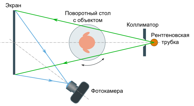
Operating principle
The installation diagram is shown in the figure below. An X-ray tube with a collimator forming a tapered beam shines through the object. X-rays that pass through the object create an image on a fluorescent screen, which is photographed by a digital camera. Turning an object, a series of images is obtained, which are used to further construct a voxel 3D model of the object and its entire internal structure.')
In reality, the installation looks like this:
Radiation source
The X-ray source is a tube already familiar to us from Ben’s past projects.
The tube is placed in a metal cylinder with a hole that allows it to shine only where it is needed. The power supply of the tube produces a voltage of 50 kV at a current of about 1 mA.
Screen
X-rays that pass through an object fall onto a luminescent screen, causing the latter to glow. Since different parts of an object absorb radiation in varying degrees, a picture is visible on the screen, where darker areas correspond to denser parts.
Swivel mechanism
In medical tomographs, the patient's body remains stationary, and around it a ring with an X-ray tube and detectors rotates. It is also easier to rotate the observed object without moving the rest of the installation.
The object is located on the turntable, which is driven by a stepper motor. The engine is driven by the Arduino. The same controller serves to control the shutter of the camera.
The pitch is 8 °, so a series of 45 shots is taken for 1 turn of the object.
Image processing
Tomography is not for nothing called "computer", because the main data processing is performed programmatically. In medical tomographs, specialized software is used, comparable in value to hardware. Ben managed the means at hand.First of all, with the help of Photoshop (in batch processing mode), perspective distortions caused by the camera being removed from the screen were removed.
Then it is necessary to obtain a series of parallel slices of the object from the images “through the gap”. For this purpose, algorithms based on inverse Radon transforms serve. They allow, having several projections of the integral absorption of X-rays, to restore the density distribution function inside the object.
Ben used Octave (open source Matlab) with a special library . The resulting series of slices looks like this:
Finally, the 3D Slicer package was used to visualize and transform the stack of slices into a three-dimensional model.
Video
Demonstration of a homemade tomograph:Source: https://habr.com/ru/post/165519/
All Articles