An inside view: the world around us
Dedicated to one beautiful person ...
Already from the second publication in the series “A View from the Inside”, the idea to look under the microscope was simple, objects that surround us every day and this found a response from the Habr residents . In addition, Anton Voitsekhovsky supported this idea, and under the electron microscope a variety of objects were already visited: from flowers to shadows. Therefore, this publication will be far from IT-related issues, but, firstly, I think it is useful in order to broaden my horizons, and, secondly, a drop of IT is still contained in it - all this would not have happened without such rapid development of computer technology and its application in electron microscopy.
')
Under the cut there will be a lot of pictures and quite a bit of text to clarify what is happening.
This article was published on the “Internet” blog for the purpose of accessibility for the entire Internet ...
Foreword
There's a lot of space down there ...
Richard Feynman, 1959.
All photographs were obtained using a scanning (also referred to as a raster) electron microscope from samples without any sample preparation, that is, without deposition of a conductive coating on the samples. Therefore, I can attend some artifacts on micrographs. For example, charging provokes distortion of straight sections in the image and does not allow to shoot at high magnifications, charged areas are usually lighter, etc. However, in my opinion, such a shooting is much more interesting from the point of view of practice. So for a rose it was possible to observe “blowing off” cells damaged by an electron beam, whereas this would not be possible if the sample were “dusty” with gold.
"Bytovuha"
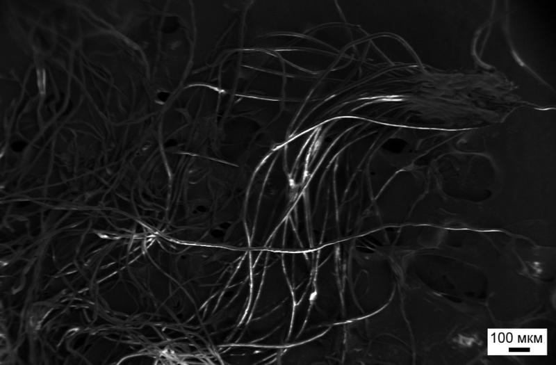
Let's start with a simple and, I think, a little less interesting. They come in different qualities, different colors and designs, but we use them almost every day. Meet the usual napkin:

The micrograph shows individual fibers, which are intertwined with each other and thus form a single mass.
Something looks like a napkin on a napkin, though the latter consists of “strings”:
Networks ...
Now a little harder - the razor of a well-known manufacturer (so that it does not look like a clear advertisement, I can reveal the brand in the comments):
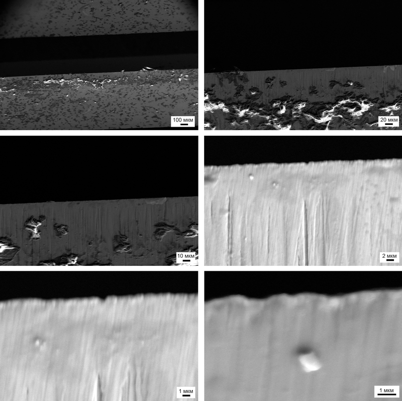
A razor blade and pieces of leather on it ...
Since there was no new blade at that time, the photos were taken from a fairly well-used machine. The photographs show clearly visible skin particles. Yes, dear ladies and gentlemen, this is what is accumulated in the form of a light, barely noticeable white residue on your razor (maybe after that everyone will stop shaving?). By the way, on the images of the edge of one of the blades, I think there is a coating. It is said that the blade itself is covered with a thin layer of hydrophobic substance, which reduces the coefficient of friction and, accordingly, irritation.
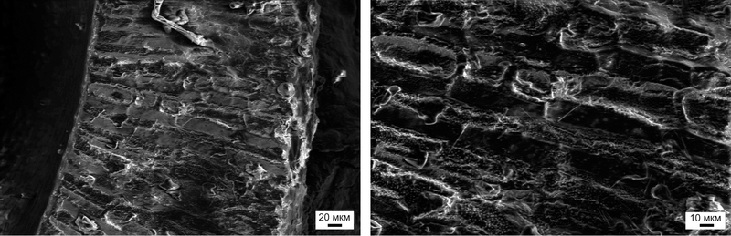
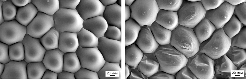
Of course, when scientific and technical progress did not reach our kitchens, many of us, when dealing with gas stoves, used matches. Someone used them not quite as intended, for example, burned a match and ate the salty residue of a match head. But not many people imagined that in this way they would actually eat “nanotechnologies” (nanoparticles that are formed after combustion).
Micrographs of a match head before (left) and after (right) ignition
I hope that among all of us there are many representatives of the beautiful half of humanity who often use the shadows, often, of course, for the mercenary purposes of attracting the attention of the strong half. However, I don’t think that cute girls wondered how, at the micro level, what they like to put on their face looks like:

These "pancakes" girls put on their face every morning to wash off in the evening
Meanwhile, the shadows consist of individual scales, with a thickness of 100 nm, which is easy to see:

That's all for now with the household part, and now let's move from the world of "inanimate" objects to the world of our favorite flowers and animals.
Plants
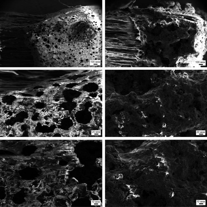
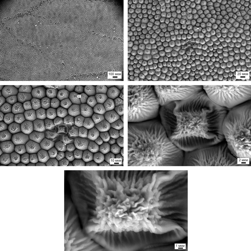
Someone swallows them together with grapes, someone diligently “catches” them and separates them from the pulp, but perhaps nobody has seen them from such a viewpoint - grape seeds:

Embryo inside the grape seed
Inside the seed kingdom: huge spherical cells waiting in the wings to begin to divide, multiply and, eventually, turn into a vine with a new bunch of grapes. But for this they need reliable protection in the form of a kind of shell, the "shell":
Notice that the cells of the envelope are coarser: they are elongated perpendicular to the surface of the shell and protrude out of the bone with their sharp corners, like guards ready to open the way for the inside to create new life with a favorable combination of circumstances:
Guardian of the seed enclosed within the grape seed
Does anyone remember what a violet looks like ?! These are such blue flowers, slightly wooly and from that very, very soft. And all because on the surface some cells allowed growths:
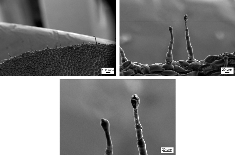
A married couple on the surface of a violet flower clarifies the relationship ...
And the rest of the surface - countless ordinary cells:
In general, the conditions for organic objects inside an electron microscope are not the best: high vacuum, drying everything without residue, a strong electric field and local heating under an electron beam. The latter, by the way, well demonstrates the bottom right picture. While I was “induced” (customized focus, astigmatism), that is, the electron beam ran through the same rather small surface area many times, it hurt the cell ...
Although without this, due to a combination of factors, biological objects inside an electron microscope do not last long:
Completely healthy, alive (left) and damaged, dying (right) cells
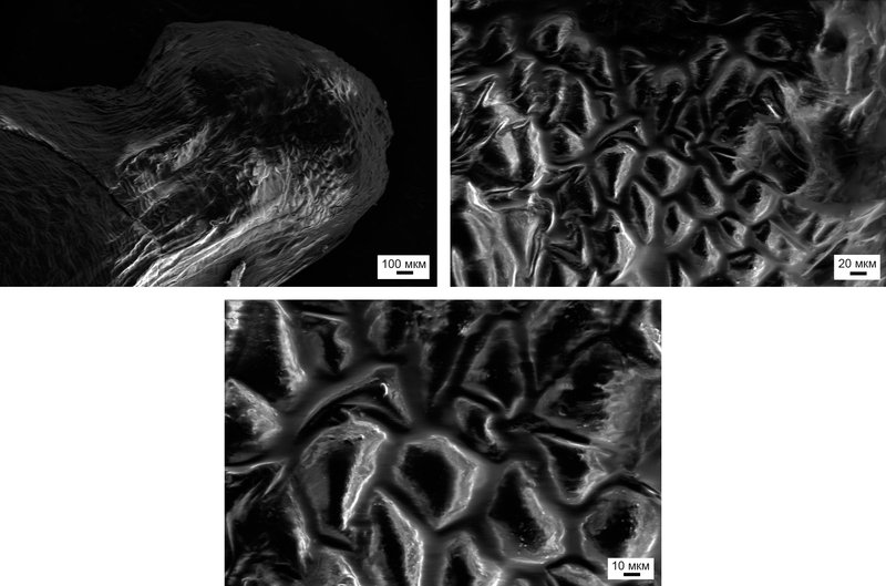
Perhaps, it was worthwhile to place these microphotographs somewhere at the beginning, for it was from them, or rather from a joke: “Let's take a rose!” - all the works presented here began:
Rose petal: looking for answers outside, and answers inside ...
The rose is just beautiful. She is a rare example of a combination of inner and outer beauty. See how strikingly different the surface of violets and roses. In violet - smooth cells, and roses - rough. One interesting property of a rose leaf is connected with this. If the violet (I think, because of its adaptability to the high-mountainous and temperate climate) the surface of the flower "collects" water, the rose pushes it away, clearing the surface. And these roughness plays a key role in this behavior of the rose flower. The air is retained in the folds, creating additional water-air interfaces, making the surface more hydrophobic than it actually is. This phenomenon has been called the Lotus effect , which is now finding more and more widespread use (for example, self-cleaning paints, car windows that are not fogged up, etc.).
And here is a gerbera with beautiful large and bright flowers:
Modest and unassuming gerbera
And inside - the very modesty. It’s not even interesting to discuss something, but let's compare ...
A gerbera with a “winter” aster (as it was presented to me, but, I suppose, it is more correct to call it an alpine aster). It seems that the petals of flowers are similar, and the structure of flowers resembles each other (after all, they belong to the same family), but in fact:
Astra seems more like a rose than its closest relative - a gerbera
The only similarity is the elongated cells. I would even say that the aster looks more like a rose flower, only the cells are more elongated along the petal itself. In addition, the place of attachment of such a petal to the inflorescence itself looks intriguing:
And then there are smooth cells. Agree, funny ?!
And I think that these "hooks" - the cells protruding from the stalks - are given by Nature to this plant is not casual, can someone think of for what?
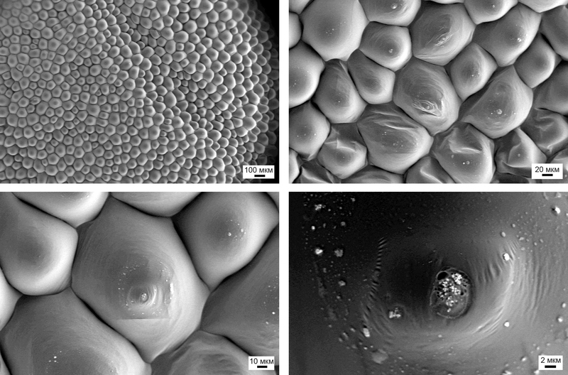
The next houseplant should love the weaker half of the audience - spathiphyllum or in the common people "Female happiness".

It seems to be nothing interesting. But the respiratory pores in the flower are absolutely amazing (nervous and impressionable, and also it is better to miss this moment for children):
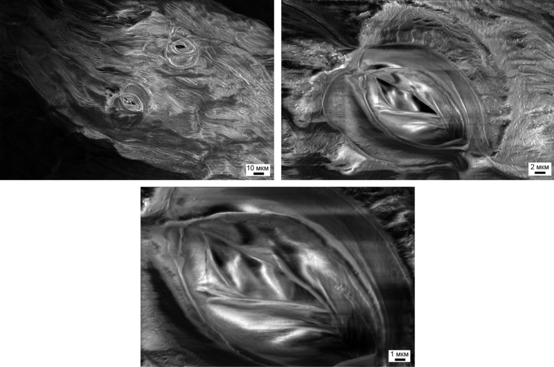
Respiratory pores
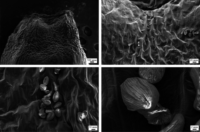
Since I caught the plant by surprise: it just wanted to shed its flower along with the seeds. By the way, it is possible that the garbage in the previous images is due to the dying of the flower and the beginning decomposition, that is, the devouring of the flower by various microorganisms. So, the seeds, but rather one single "seed"
Spathiphyllum seed - all around the cell
I don’t even know what these small particles found in the place of attachment of the seed to the plant belong to (can there be any pollen residues ?!):
And finally, a little about animals, but rather about their external cover - hair and feathers ...
Animals
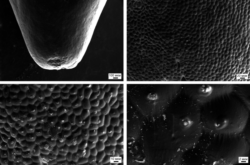
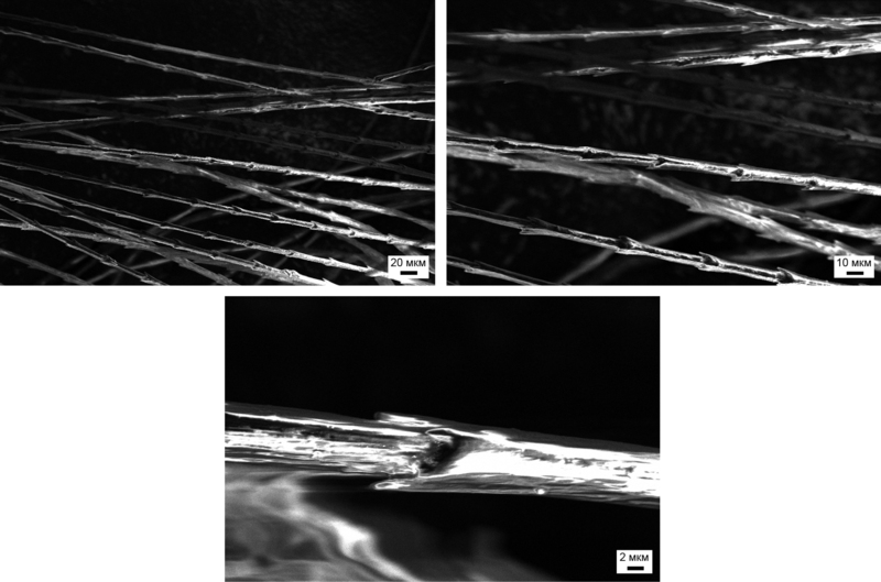
Let's start with some trivial - human hair (had to be sacrificed from a beard):
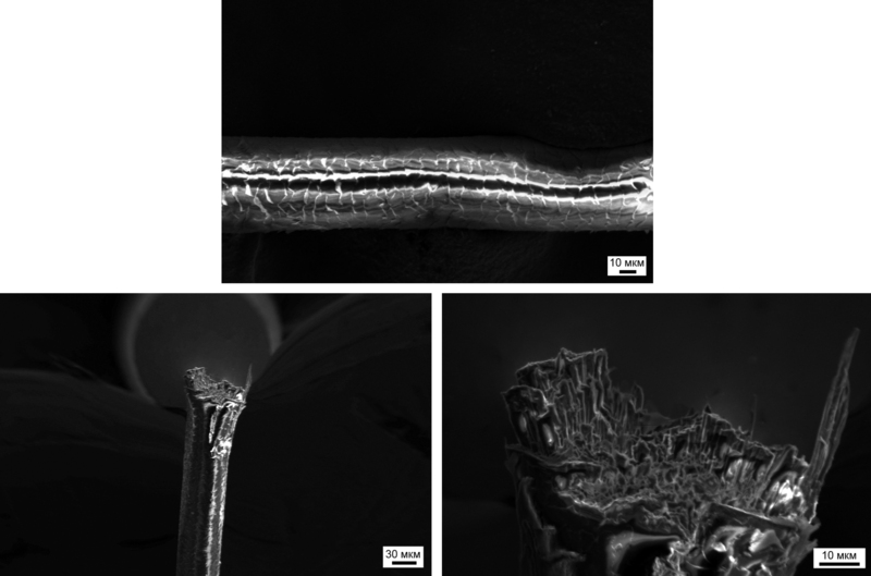
The hair didn’t fit completely into the place allotted to him, I had to cut it ...
Of course, we all, at a minimum, saw in the advertisement that the hair has a scaly structure (in the upper photo you can see it well), but absolutely few saw the hair in the section ... Our hair looks like this or so after going to the hairdresser. If it is interesting, then you can look at the parts of the human body in color here - many beautiful SAM micro-micrographs.
One of my friends decided to start a budgerigar , and when I came to her to show photos of the rosette, I was immediately handed a feather, just thrown out of my body by the parrot either from the wing or from the tail. But this is understandable, pay attention to the number of microorganisms and other filth in the down:
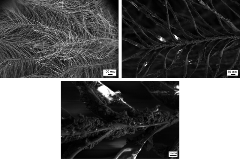
Micrographs of the budgie down close to the base of the pen
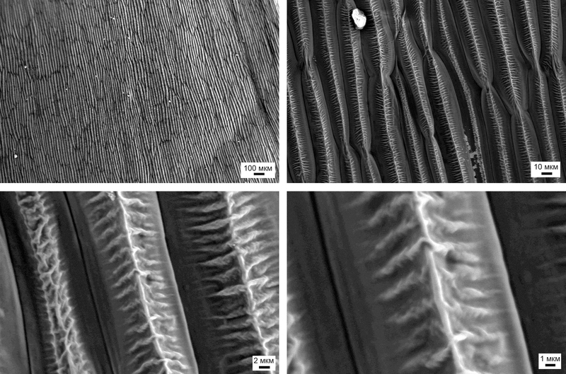
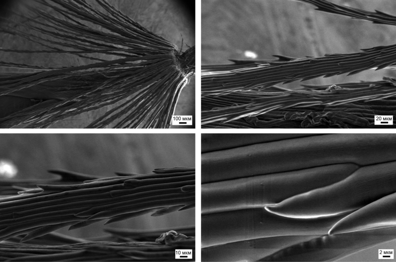
Although the main part of the pen (ostevaya) is preserved in excellent form. See how perfectly “hairs” are laid in the feathers - this is all so that the birds can fly and maneuver in the air, so that their wings are not a sieve for the air, and, of course, this also has its aesthetic function:
The rest of the pen wavy parrot
Well, when one of the researchers at our laboratory at Moscow State University found out about these photos, a small peacock feather was given to me to be torn apart. Apparently, it once belonged to a young individual, since it was no more than 10–15 cm in length.
Let's start, as in the case of the parrot, with the "undercoat" in which microorganisms also live. Unfortunately, at that moment Wojciechowski was not there, eager to see the running "green men" on the surface of biological samples. Here they, however, do not run, but simply stuck to the hairs of fluff:
And also a few micrographs of down, more precisely that part of the hairs, which is removed from the core of the pen:
Like the "hairs" are inserted one into the other.
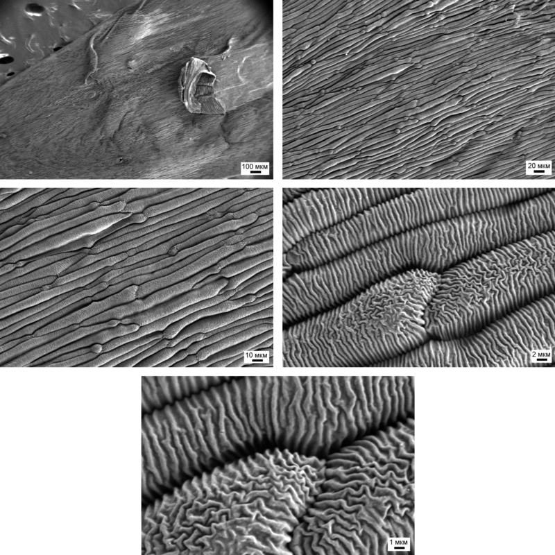
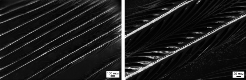
The main part of the pen looks like this:
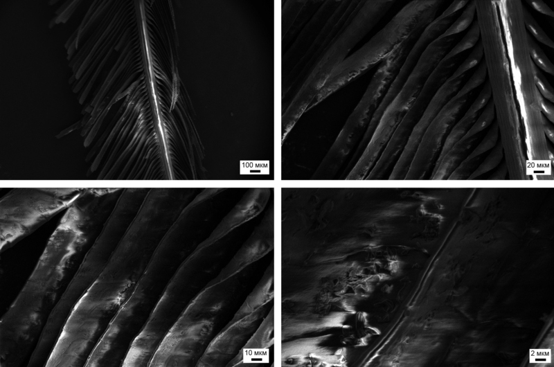
Here, wide flattened “hairs” are stacked on each other overlapping, which ensures monolithic feathers, lower air permeability and its beauty, both in battles between males and in relationships with members of the opposite sex.
But that's not all. If we compare it with a wavy parrot, whose hairs are short (about 100 microns in length) and rigid and do not have links with each other, then in the peacock feather such hairs are longer (up to 300-500 microns in length) and, accordingly, less rigid . And so that they do not mess up when wearing such a chic feather costume, Nature has come up with the following:
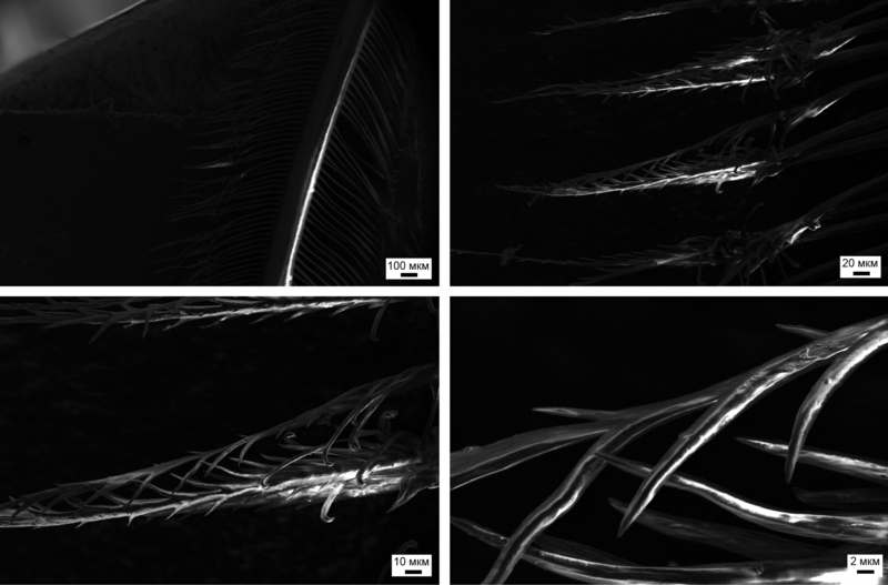
The progenitor of zip-lock, created by Nature
Separate hairs are linked at the ends between themselves and with the help of such devices can link with adjacent feathers, etc.
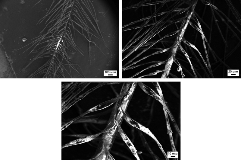
Finally, the hairs inside the feather have an interesting structure with broadening and thinning along one hair. This determines the iridescent color, the play of colors and the peacock feather in the sun:
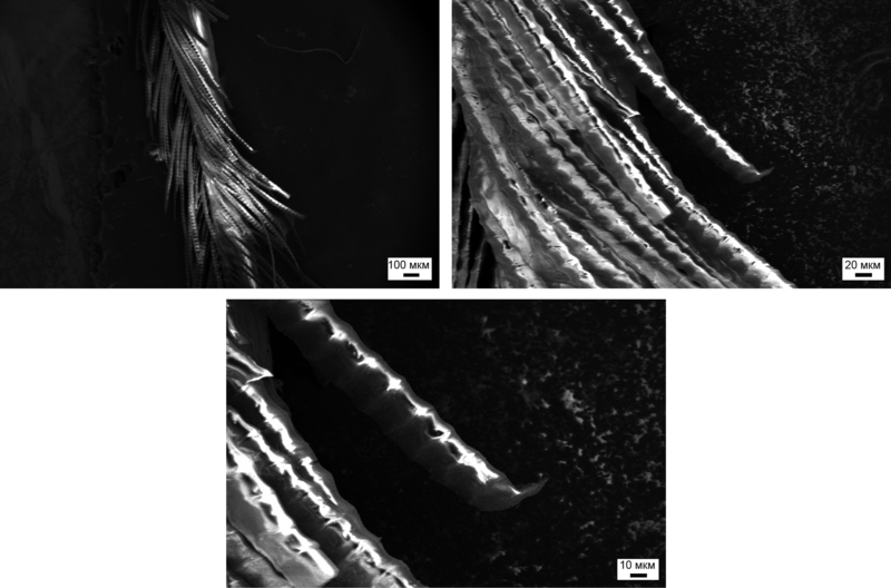
It is precisely such thickenings of individual hairs that are responsible for the play of the light of the peacock feather.
You can read about the structure of feathers here .
Afterword
At the moment I have everything. If you have ideas that you could see in the electron microscope, saw, what else to write on Habré, feel free to send me your wishes in any available way (all contacts are in the profile) or to smirnovea_ (at) _gmail .com (without the “_” signs, and (at) - well, you yourself know ...).
PS: OSRAM movie was able to fasten the subtitles for the New Year holidays, enjoy here . And do not forget to subscribe to the channel , I hope, there will soon be a “home-grown” movie about plasma in the installation for spraying conductive coatings, which was not so good seen in this video ...
PPS: If someone has not read the previous articles, you can see them here:
Opening the Nvidia 8600M GT chip , a more detailed article is given here: Modern chips - a view from the inside
An inside look: CD and HDD
An inside look: LED bulbs
An inside look: the LED industry in Russia
An inside look: Flash and RAM
Object list for the future
Interesting:
Displays (e-ink, conventional TFT with a resistive matrix). If someone donates a touch capacitive or Gorilla Glass, I will be very grateful ...
Cut the cage and gently look at the insides
In addition to the match - chirkash
Electrolytic capacitor
Optical fiber
floppy disks, magnetic tapes, vinyl records, film, films (only the samples are needed)
The surface of scratch cards or instant lottery tickets
The surface of "normal" polished plastic (PVC, PP, etc.) and the surface of plastic, covered with "soft touch" (again, need samples)
The edge of the metal line
Ball Pen / Ball Pens
Braid and the inside of the guitar string (you need to take a new and heavily used, but in general the guitar string is here )
meat / fish and other edible things
Radio-frequency tags (RFID) - just compare the usual RosNanovskaya super-duper tag with some usual
Skepticism:
Holographic sticker (the physical principle of Holography hints that it will be just a monolithic piece of polymer, but as soon as the opportunity arises, I will see)
Tap water, beer, other liquids (sterilization kills everyone, but it's easy to see nanoparticles)
The surface and the interior of porous materials such as polyethylene foam, polypropylene foam, polyurethane foam, filter from cigarettes (I even watched the earplugs straightened into an optical microscope - was not impressed, but I would like to play with an electron microscope)
Already have on the Internet:
Dust mites and sperm (like discussed in the comments - look here and here )
First , the full list of published articles on Habré:
Opening the Nvidia 8600M GT chip , a more detailed article is given here: Modern chips - a view from the inside
An inside look: CD and HDD
An inside look: LED bulbs
An inside look: the LED industry in Russia
An inside look: Flash and RAM
An inside view: the world around us
An inside look: LCD and E-Ink displays
An inside look: matrix digital cameras
An inside look: Plastic Logic
An inside look: RFID and other tags
An inside look: graduate school at EPFL. Part 1
An inside look: graduate school at EPFL. Part 2
An inside view: the world around us - 2
An inside view: the world around us - 3
An inside look: the world around us - 4
An inside look: 13 LED lamps and a bottle of rum. Part 1
An inside look: 13 LED lamps and a bottle of rum. Part 2
An inside look: 13 LED lamps and a bottle of rum. Part 3
An inside look: IKEA LED strikes back
An inside view: are Filament lamps good for you?
and 3DNews:
Microview: a comparison of modern smartphones displays
Secondly , in addition to the blog on HabraHabr , articles and videos can be read and viewed on Nanometer.ru , YouTube , and Dirty .
Thirdly , if you, dear reader, liked the article or you want to stimulate writing new ones, then act according to the following maxim: “pay what you want”
Yandex.Money 41001234893231
WebMoney (R296920395341 or Z333281944680)
Sometimes briefly, and sometimes not so much about the news of science and technology, you can read on my Telegram channel - welcome;)
Source: https://habr.com/ru/post/136234/
All Articles